Original Article – DOI: 10.33594/000000899
CPB (60): 616 - 645
Accepted: 23.10.2026 - Published: 13.11.2026
Cellular and Molecular Mechanisms of Wound Healing and Therapeutics Innovations
bDepartment of Medical Laboratory Technology, Faculty of Applied Medical Sciences, University of Tabuk, Tabuk 71491, Saudi Arabia,
cIndo-American Cancer Research Foundation, Basavatarakam Indo-American Cancer Hospital and Research Institute, Banjara Hills, Hyderabad 500034, India,
dMultidisciplinary Research Unit (MRU), Government Medical College (GMC), Srinagar 190010, Jammu & Kashmir, India,
eZamzam Cancer Research Institute, Zamzam Medical Institute, Ganderbal, J and K, India-191201
Keywords
Abstract
Chronic wounds are a significant and growing global clinical problem owing to increasing rates of diabetes, vascular diseases, ageing, and metabolic disorders. In contrast to acute wounds, which follow a coordinated sequence of haemostasis, inflammation, proliferation and remodelling, chronic wounds fail to progress through the healing process, leading to delayed or unfinished recovery. There is mounting evidence that this malfunction lies primarily in dysregulated intracellular signalling and is not necessarily in a lack of growth factors or cellular components. There is prolonged stimulation of pro-inflammatory pathways, disrupted angiogenic and regenerative signalling, excessive extracellular matrix degradation, cellular senescence and impaired immune resolution in chronic wound environments. The most important signalling networks, such as TGF-b/Smad, PI3K/Akt/mTOR, MAPK, NF-kB, Wnt/b-catenin, JAK/STAT, Hippo-YAP/TAZ, Notch, and hypoxia-inducible factor-1a, are crucial in the coordination of wound repair, but are functionally impaired in chronic pathological diseases. The pathways demonstrate a widespread crosstalk, forming a self-maintaining feedback loop that drives inflammation, oxidative stress, endothelial dysfunction, and a lack of cellular responsiveness. This review provides a detailed description of the physiology of normal wound healing, compares acute and chronic wound pathology, and critically discusses the molecular signalling defects that promote the persistence of chronic wounds. Additionally, it identifies emerging therapeutic approaches aimed at regulating dysregulated signalling networks, such as small-molecule modulators, biologics, biomaterials, gene- and cell-based therapies and exosome-mediated therapies.
Introduction
Chronic wound healing represents a failure of the skin’s intrinsic regenerative capacity and remains a major unresolved problem in modern medicine (1). In physiological conditions, cutaneous wounds are repaired in a highly regulated series of cellular and molecular processes that restore tissue integrity and barrier functionality. This coordinated programme, however, fails in chronic wounds, and leads to prolonged wound persistence (months to years) despite standard clinical care (2). The wound repair process is centred on a system of intracellular signalling pathways that regulate inflammation, cell migration, cell proliferation, angiogenesis and extracellular matrix (ECM) turnover (3). These are activated by growth factors, cytokines, mechanical cues, and metabolically activated pathways, and work in a strictly orchestrated time order. This temporal regulation is lost in chronic wounds (4). Instead, pro-inflammatory and stress-activated pathways remain persistently engaged, while regenerative signalling becomes blunted or ineffective (5). This imbalance causes defects in the migration of keratinocytes, fibroblast dysfunction, endothelial cell impairment, and immune dysregulation. Systemic diseases, including diabetes mellitus, vascular insufficiency, obesity, and old age are most often related to chronic wounds. These conditions cause metabolic stress, ischemia and immune dysfunction, which work together to change the intracellular signal transduction of the wound-resident cells (6). Experimental and clinical data show that growth factors are often present in chronic wounds without triggering downstream signalling, because of receptor desensitisation, oxidative damage, and epigenetic modifications (7). This understanding has changed therapeutic approaches to no longer focus on growth factor supplementation, but to focus on modulating signalling pathways in a targeted manner. The recent developments in molecular biology indicate that a range of signalling axes, such as TGF-b/Smad, PI3K/Akt/mTOR, MAPK, NF-kB, Wnt/b-catenin, and hypoxia-inducible pathways, have been identified as key regulators of the wound healing outcomes (4). The disruption of these pathways does not happen in isolation, and instead of competing, they interact to form feedback loops which reinforces long-term inflammation and tissue destruction. This mechanistically based approach of repairing healing competence involves targeting signalling pathways (8). Pathway-based interventions can overcome the issue of therapeutic resistance in chronic wounds by correcting intracellular communication, instead of focusing on the surface-level pathology (9). The current review is concerned with the physiological foundations of wound healing, the molecular pathways of chronic wound pathology, and novel therapeutic approaches that are aimed at dysregulated signalling networks specifically. Rather than treating these nine signalling axes as independently dysregulated pathways, the organising concept applied throughout this review is that chronic wounds represent a self-stabilising, network-level signalling state, in which crosstalk between pathways — rather than dysfunction confined to any single pathway — sustains the non-healing phenotype. This crosstalk-centred framework is used to interpret pathway-specific findings within an integrated context in the sections that follow, and provides the rationale, developed later in this review, for combination rather than single-target therapeutic strategies.
Overview of Wound Healing
Wound healing is an evolutionarily conserved biological process that is aimed at the rapid restoration of tissue continuity after injury (10). Physiological descriptions frame wound healing as a dynamic process rather than a fixed sequence of events, with several phases occurring simultaneously and overlapping over time. The different phases are controlled by distinct yet interrelated signalling pathways that coordinate tissue repair while limiting inflammation and fibrosis. This process begins immediately after injury, with vascular and cellular reactions that restrict blood loss and activate immune surveillance (11). In addition to structural components, platelets, immune cells, stromal cells and epithelial cells release soluble mediators that activate intracellular signalling cascades. These cues coordinate cell migration, proliferation, differentiation, and recruitment. Effective healing relies on the capacity of cells to perceive the environmental signals and convert them into suitable transcriptional and metabolic reactions (12). Intercellular communication is the key to wound healing. Growth factors, including platelet-derived growth factor (PDGF), transforming growth factor-b (TGF-b), vascular endothelial growth factor (VEGF), and fibroblast growth factors (FGFs) can stimulate receptor-mediated signalling in target cells. All these signals converge onto intracellular pathways such as PI3K/Akt, MAPK and Smad signalling which orchestrate gene expression programs required for tissue repair (13-17). Any disruption within this signalling hierarchy may disturb healing. Immune cells have a dual purpose in the healing process of wounds; as they both protect against infection and regulate tissue repair. In particular, macrophages combine inflammatory and repair signalling through the production of cytokines, growth factors and matrix-remodelling enzymes (18). The transition of inflammation to tissue reconstruction depends on their functional plasticity. This transition failure is a characteristic of chronic wounds (19). The microenvironment of the wound is also significant. Signal transduction depends on oxygen tension, mechanical stress, extracellular matrix composition and metabolic status. An example of this is hypoxia that temporarily switches on angiogenic signalling by HIF-1a, but in the long term, hypoxia upsets cellular metabolism and signal fidelity (20). The literature highlights the fact wound healing is not coupled to the individual molecular events but the systems-level coordination of signalling networks (21). Overall, wound healing is a well-coordinated biological programme that only functions correctly upon tight control of wound healing signalling. This normal framework is critical in determining the causes and effects of failure of healing in chronic wounds.
Acute vs. Chronic Wounds
Wounds are generally distinguished into acute and chronic wounds based on the time of the healing process and the biological behaviour during the healing process. Even though both are similar in their physiological stages of repair, their healing processes, the pathophysiological processes, and clinical changes vary considerably (22). Acute wounds are those that follow a systematic, sequential healing process and fully restore tissue integrity. Examples include surgical lacerations, traumatic lacerations, burns and abrasions. The time required for these wounds to heal, typically days to weeks, depends on wound characteristics and the individual's health status (23). Acute wound healing has four coordinated stages that include haemostasis, inflammation, proliferation, and remodelling. Immediately after injury, haemostasis prevents blood loss through clot formation. This is followed by an inflammatory stage that is characterized by the migration of immune cells, including neutrophils and macrophages that destroy debris and inhibit infection. There is fibroblast migration, angiogenesis, re-epithelialization and extracellular matrix (ECM) deposition in the proliferative phase. Lastly, the tissue is reinforced in the remodelling stage by reorganizing collagen and contracting of the wound (22). Acute wounds have a well-regulated inflammation which heals effectively and leads to a progression of the tissue regeneration process. VEGF, platelet-derived growth factor (PDGF) and transforming growth factor-b (TGF-b) are growth factors that are instrumental in cell proliferation and angiogenesis (24). When the same processes continue without a pause, the healing process of the body is characterized by low scarring and restoration of functions. Nevertheless, infection, lack of oxygen, malnutrition or systemic illnesses may affect the result of acute wounds. In case these conditions are not changed, acute wounds cannot heal in a proper way and develop into chronic ones (25). Chronic wounds are defined by their failure to progress through the typical stages of healing and are usually unhealed after 4–6 weeks. They include diabetic foot ulcers, venous leg ulcers, pressure injuries, and arterial ulcers. These wounds constitute a major clinical challenge because of their long healing time, high susceptibility to infection, high recurrence rate, and high healthcare expenditure (26). Inflammation in chronic wounds is dysregulated and prolonged. Unlike acute wounds, chronic wounds remain in the inflammatory phase, resulting in an overproduction of pro-inflammatory cytokines, proteases, and reactive oxygen species (ROS) (27). This chronic inflammation destroys surrounding tissue and impairs growth factor signalling and fibroblast and keratinocyte function. Impaired angiogenesis is one of the significant factors contributing to chronicity: reduced blood circulation and oxygen supply limit nutrient delivery, slowing granulation tissue formation and epithelial regeneration (28). Hypoxia which is prevalent in diabetic wounds as well as ischemic wounds further damage the tissues and prevent cellular growth. Microbial colonization and biofilm formation are additional characteristics of chronic wounds. The bacteria in biofilm remain resistant to immune clearance and antimicrobial therapy to maintain low-grade infection and inflammation. Biofilms disrupt re-epithelialization and play a major role in impaired wound healing (29). Also, in chronic wound healing, there is a change in the extracellular matrix remodelling. The overactivity of the matrix metalloproteinases (MMPs) in comparison to reduced levels of their inhibitors contributes to the breakdown of collagen and growth factors (30). Such imbalance is not conducive to the reconstruction of the tissues and instead promotes tissue breakdown rather than repair. Unlike acute wounds, which progress through tightly regulated and overlapping phases of repair, chronic wounds remain trapped in a state of unresolved inflammation with impaired proliferation and remodelling (Fig. 1).
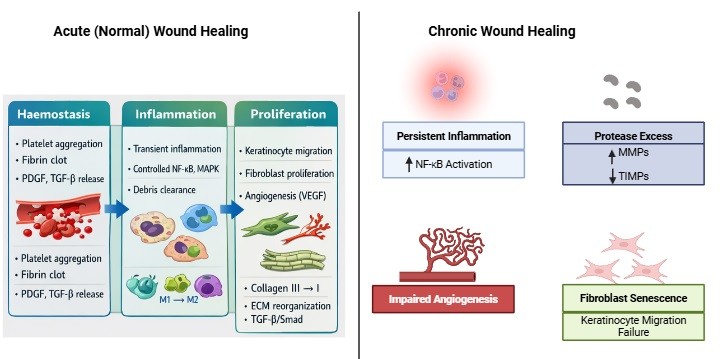
Fig. 1: Acute vs. Chronic Wound Healing. Left panel (Acute Wound Healing): Sequential stages of haemostasis (platelet aggregation, fibrin clot, PDGF/TGF-β release), inflammation (transient NF-κB/MAPK signaling, M1-to-M2 macrophage transition, debris clearance), and proliferation (keratinocyte migration, fibroblast proliferation, VEGF-mediated angiogenesis, collagen remodeling, ECM reorganization, TGF-β/Smad activation). Right panel (Chronic Wound Healing): Persistent inflammation (sustained NF-κB), protease excess (MMP/TIMP imbalance), impaired angiogenesis, fibroblast senescence, and keratinocyte migration failure, leading to delayed or non-healing wounds.
3.1 Stages of Wound Healing
3.1.1 Haemostasis Haemostasis is an immediate response to tissue injury that serves protective and signalling functions in wound healing. Exposed tissue factors and collagen on disruption of the vascular system cause platelet adhesion, aggregation, and clotting. Although the main governing principle in haemostasis is to avoid excessive bleeding, this step also forms the biochemical basis of the further healing events (31). On activation, platelets secrete a focused set of bioactive mediators, including platelet-derived growth factor (PDGF), transforming growth factor-β (TGF-β), fibrinogen, and fibronectin, which become incorporated into the fibrin clot that serves as a temporary extracellular matrix. This scaffold provides mechanical stability and spatial cues that guide the infiltration of inflammatory cells and migrating stromal cells. The fibrin matrix is not merely a passive scaffold, however: it actively shapes cell behaviour by exposing ligands that engage integrins and growth factor receptors (32). On a molecular level, haemostasis triggers early cellular signalling cascades in cells in the immediate environment. Platelet growth factors and receptor tyrosine kinases on fibroblasts, endothelial and keratinocytes prime intracellular signalling pathways including PI3K/Akt and MAPK (33). These initial indications do not cause proliferation at all but rather they equip cells to become responsive in the future by stimulating gene expression and metabolic activity. Excessive persistence of clots may hinder cell migration, and accelerated breakdown of clots may impair structure. Maintaining a balanced fibrinolysis ensures the timely removal of the provisional matrix and this is substituted by newly generated extracellular matrix in later stages (34). During the normal healing process, haemostasis is completed rapidly, usually in hours to a day. Any failure during this phase, including platelet dysfunction or clot defects, may slow subsequent healing. Therefore, haemostasis is an important signalling crossroads that combines vascular injury and cellular repair (35).
3.1.2 Inflammatory Phase The inflammatory stage is the immune-surveillance and wound-cleansing phase of tissue repair. After haemostasis, vascular permeability increases, and immune cells enter the site of the wound. The initial responders are neutrophils, which quickly clear debris and pathogens by means of phagocytosis and release of antimicrobial agents. Although their activity is critical in infection prevention, as an additional point, it is important to note that neutrophil activity should be transient to avoid collateral tissue damage (36). Macrophages subsequently become a major immune-cell population and key regulators of this stage. Macrophages arise in part from circulating monocytes and combine wound microenvironmental signals, as well as organize immune and repair functions. They release pro-inflammatory cytokines like TNF-a and IL-1b to maintain early defence, and at the same time release growth factors to trigger tissue repair (37). At the signalling level, transcriptional programmes controlled by NF-kB and stress-responsive MAPK are triggered by the inflammatory cells. These pathways regulate the production of cytokines, chemokine gradients and the production of proteases. When normal healing occurs, the activation of these pathways is strong yet self-regulated. Inflammatory signals gradually repress the inflammatory gene expression, which progresses to the proliferative phase (38). The inflammatory phase is also critical for extracellular matrix turnover. The release of matrix metalloproteinases by the immune cells breaks the damaged components of the matrix and creates space where new tissues are formed (39). This degradation must be tightly regulated; excessive protease activity can destroy growth factors and interfere with cell adhesion. The research here points to a broader principle: inflammation itself is not necessarily detrimental to healing. Rather, it is the failure to resolve inflammation that drives pathological outcomes. During normal wound healing, inflammatory signalling is dampened as macrophages assume a reparative phenotype and immune cell density declines (40).
3.1.3 Proliferative Phase The proliferative phase marks the transition from immune-mediated defence to tissue reconstruction. At this phase, the fibroblasts, keratinocytes and endothelial cells are very active and they restore the wound by synchronizing cellular growth and matrix deposition. This stage is mentioned in the literature as the most metabolically stressful phase of healing, which entails a prolonged growth factor signalling and the provision of sufficient nutrients (41). Fibroblasts move into the wound bed and express different elements of the extracellular matrix, such as collagen type III, proteoglycans, and glycosaminoglycans. These elements constitute granulation tissue which substitutes the provisional fibrin matrix. Signalling via growth factors (PDGF, TGF-b and FGF receptors) stimulates intracellular signalling pathways including PI3K/Akt and ERK which stimulate fibroblast proliferation and survival (42). At the same time, the keratinocytes at the edge of the wound experience phenotypic changes, which allow them to move across the surface of the wound. It is a process called re-epithelialization and entails the reorganization of cytoskeleton, integrin switching and inhibition of terminal differentiation. At this stage, Wnt/b-catenin and MAPK signalling pathways are important in controlling the motility of the keratinocytes and gene expression (43). The other characteristic of proliferation is angiogenesis. The response to hypoxia and growth factors like VEGF by endothelial cells occurs by developing new capillary networks in the granulation tissue. This neovascularization provides perfusion of oxygen and nourishment, which promotes further growth of the tissues (44). The proliferative stage is very sensitive to disruptions in signalling. Poor responsiveness to growth factors, or angiogenic signalling or a metabolic stress can delay healing at this stage, which is common in chronic wounds (45).
3.1.4 Remodelling and Maturation Phase The remodelling phase is the final phase of wound healing, when the newly developed tissue is reorganized to achieve the restoration of mechanical strength and functional integrity. Compared to previous stages, remodelling is also less cellular and less metabolic (46). The collagen type III that is deposited during proliferation is replaced by collagen type I, which is stronger and is thus built up during remodelling, which causes fibroblasts to develop into myofibroblasts, which produce a contractile force that decreases the size of the wound. TGF-b and mechanical signalling pathways at the molecular level mediate the activity of myofibroblasts and the reorganization of matrix (47). The activity of matrix metalloproteinases and inhibitors regulate the dismantling and the re-assembly of the extra-cellular matrix components. This balance will provide adequate orientation of collagen fibers along mechanical stress lines that increase tensile strength. The high deposition of the matrix or long-term action of myofibroblasts may result in fibrosis and hypertrophic scarring (48). Apoptosis plays a crucial role during the remodelling phase. The surplus endothelial cells, fibroblasts and immune cells undergo apoptosis after fulfilling their functions. Inability of this clearance process creates abnormal tissue architecture (49). Signalling pathways slowly resume normal levels in the process of successful healing to restore tissue homeostasis. The remodelling period therefore ensures that wound healing progresses toward long-term tissue stability as opposed to continuous activation (50). The sequential phases of normal wound healing and their associated cellular events and dominant signalling pathways are summarized in Table 1.
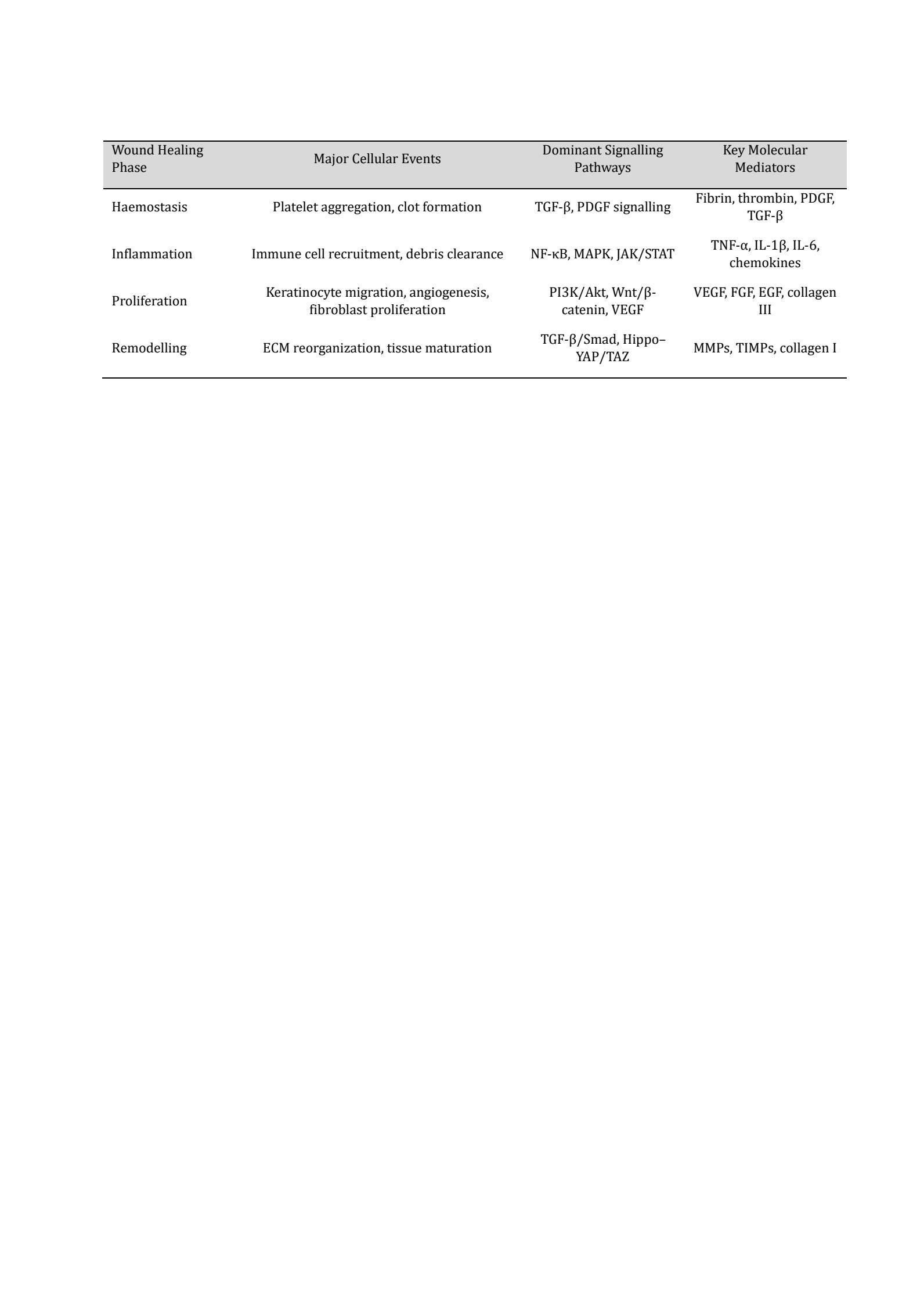
Table 1: Phases of Normal Wound Healing and Key Molecular Signalling Events
4. Global Burden and Clinical Challenges of Chronic Wounds Chronic wounds represent one of the most persistent and underestimated public health challenges worldwide. Their burden extends far beyond localized tissue damage, affecting patients physically, psychologically, socially, and economically. Globally, chronic wounds affect millions of individuals, with prevalence estimates suggesting that approximately 2-3 per 1, 000 people suffer from a chronic non-healing wound at any given time (51). This burden is expected to rise substantially in the coming decades due to population aging, increasing prevalence of diabetes, obesity, cardiovascular diseases, and prolonged life expectancy. Among the most commonly encountered chronic wounds are diabetic foot ulcers, venous leg ulcers, arterial ulcers, and pressure injuries, each contributing significantly to morbidity and healthcare utilization (52). The global burden of chronic wounds is particularly evident in elderly populations, who are disproportionately affected due to age-related physiological changes, reduced tissue regeneration capacity, impaired immunity, and higher prevalence of comorbidities (53). Studies indicate that chronic wounds are more prevalent in individuals over 65 years of age, with rates escalating dramatically among those with diabetes or vascular disorders. Diabetic foot ulcers alone affect millions worldwide each year and represent one of the leading causes of non-traumatic lower-limb amputations. Alarmingly, the five-year mortality rate following a diabetic foot ulcer or related amputation rivals or exceeds that of several cancers, underscoring the severity of this condition (54). Beyond clinical outcomes, chronic wounds exert a substantial economic burden on healthcare systems. The cost of managing chronic wounds includes repeated hospital visits, prolonged hospitalizations, frequent dressing changes, surgical debridement, antimicrobial therapy, advanced wound care products, and long-term rehabilitation. In many countries, wound care accounts for a significant proportion of healthcare expenditure, often comparable to or exceeding costs associated with cardiovascular diseases or cancer. The financial burden is further compounded by indirect costs such as loss of productivity, long-term disability, and the need for caregiver support. For patients and families, this often translates into reduced quality of life, emotional stress, and financial hardship (55). From a clinical perspective, chronic wounds are particularly difficult to manage because of their complex, multifactorial nature. Unlike acute wounds, which progress through an orderly and timely healing cascade, chronic wounds frequently stall in the inflammatory phase: persistent inflammation, excessive protease activity, impaired growth factor signalling, reduced angiogenesis, and poor cellular responsiveness together hinder tissue repair, compounded by ischaemia, hypoxia, and recurrent infection that further delay healing (56). The presence of biofilms—structured communities of microorganisms encased in a protective matrix poses a major clinical challenge, as these biofilms reduce the effectiveness of antimicrobial agents and contribute to treatment resistance and wound chronicity (57). Another major challenge in chronic wound management is the high prevalence of underlying systemic conditions. Diabetes mellitus, peripheral arterial disease, venous insufficiency, malnutrition, obesity, and advanced age significantly impair wound healing mechanisms. Poor glycemic control, for instance, leads to microvascular damage, neuropathy, and immune dysfunction, all of which predispose individuals to ulcer formation and delayed healing. Similarly, venous hypertension and arterial insufficiency compromise tissue oxygenation and nutrient delivery, further perpetuating non-healing wounds (58). These systemic factors often coexist, making management more complex and necessitating a comprehensive, multidisciplinary approach. Although these four wound types share overlapping downstream features such as persistent inflammation and impaired angiogenesis, their primary aetiological drivers and the signalling pathways most consistently reported to be dysregulated differ, as summarised in Table 2.
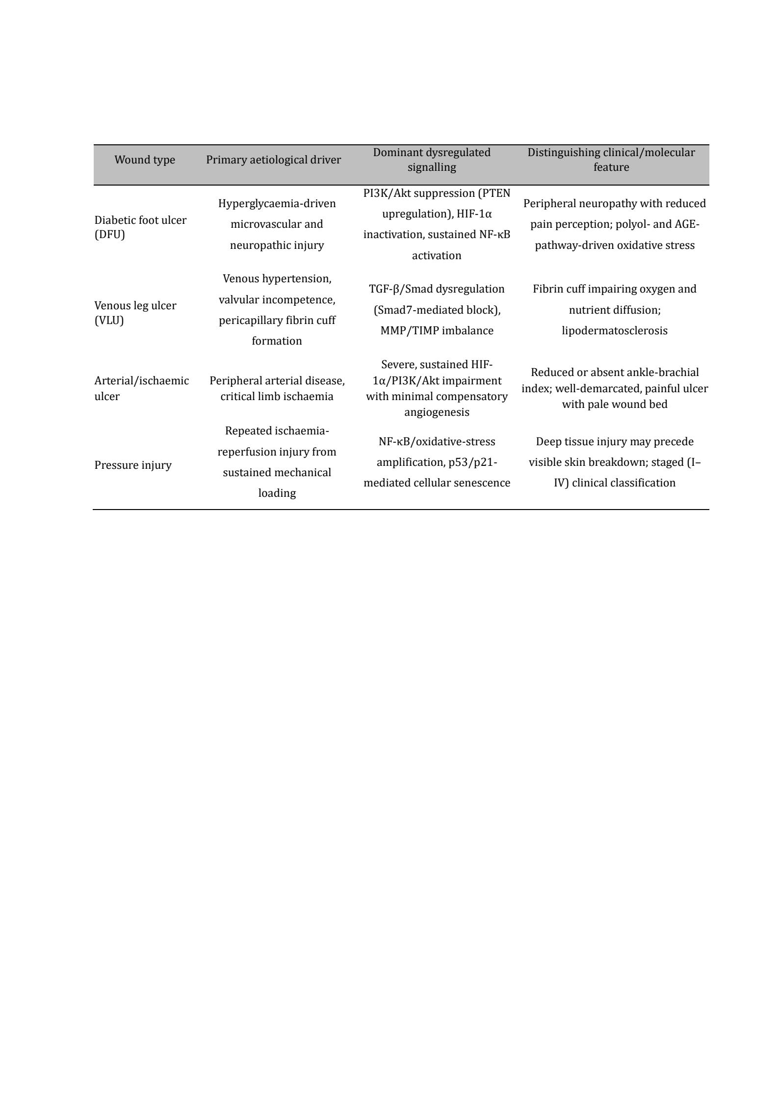
Table 2: Comparative Aetiological and Signalling Characteristics of the Principal Chronic Wound Types. Pathway-specific evidence underlying each entry is discussed, with citations, in Sections 5 and 6
5. Pathophysiology of Chronic Wounds
5.1 Persistent Inflammation The hallmark pathological feature of chronic wounds is persistent inflammation, representing a fundamental failure of the normal resolution process. In physiological wound repair, inflammatory signalling is transient and limited, modulated to debride debris and pathogens before yielding to regenerative processes (59). However, in chronic wounds, these inflammatory processes fail to resolve and instead cause ongoing tissue damage and repair failure. At the cellular level, chronic wounds are marked by sustained infiltration of neutrophils and macrophages locked in a pro-inflammatory state. Neutrophils continue releasing proteases, reactive oxygen species (ROS), and antimicrobial peptides; whereas this process is protective in acute injury, it becomes cytotoxic when persistently stimulated. These mediators degrade growth factors, damage extracellular matrix (ECM) constituents, and disrupt cell migration (60). In chronic wounds, macrophages exhibit defective phenotypic switching. In a healthy state, macrophages switch from an inflammatory to a reparative phenotype, which facilitates angiogenesis and matrix production. This transition is incomplete or delayed in chronic wounds, leading to sustained secretion of pro-inflammatory cytokines including TNF-α, IL-1β, and IL-6. This feedback enhances NF-κB and stress-activated MAPK signalling, which further supports the expression of inflammatory genes (61). Chronic inflammation also interferes with wound microenvironment. Increased protease activity causes the overexpression of ECM and cell surface receptors, which decreases the sensitivity of fibroblasts and keratinocytes to growth factors. The inflammatory cytokines also prevent the migration and proliferation of the keratinocytes, which directly interferes with re-epithelialization (62). Local wound factors are not, however, the only cause of chronic inflammation. Systemic diseases such as diabetes, obesity, and vascular disease create a pro-inflammatory environment that amplifies local immune reactions. Hyperglycaemia, advanced glycation end products, and ischaemia alter intracellular signalling thresholds, making immune cells harder to resolve (63). Sustained inflammation therefore becomes a vicious pathological cycle: inflammatory signalling causes tissue damage, and tissue damage in turn amplifies inflammation. Breaking this cycle is essential to restoring healing competence in chronic wounds.
5.2 Impaired Angiogenesis and Vasculogenesis Wound healing also requires adequate vascularization because newly created tissue needs oxygen, nutrients and immune surveillance. Angiogenesis and vasculogenesis is severely impaired in chronic wounds, which adds to ischemia and metabolic stress in the wound bed (64). Angiogenesis during normal wound healing is stimulated by hypoxia-induced signalling and release of growth factors, specifically vascular endothelial growth factor (VEGF). The endothelial cells react to such signals by multiplying, migrating and creating new capillary networks. Although backed by hypoxia, endothelial cells in chronic wounds are less responsive to the angiogenic signals (44). The activation of downstream pathways including PI3K/Akt and ERK is defective as a central contributor of this failure. There is an increase in endothelial dysfunction that is additionally induced by oxidative stress and chronic inflammation (65). Reactive oxygen species cause damage to endothelial cell membranes, receptors, and intracellular signalling proteins which affects nitric oxide bioavailability and vascular relaxation. Endothelial junctions are also perturbed by the inflammatory cytokines, which elevate the vascular permeability, but do not enhance the effective vessel formation (66). Vasculogenesis, which involves the recruitment of circulating endothelial progenitor cells, is similarly affected: persistent systemic inflammation and metabolic disease reduce the number and activity of these progenitor cells, thereby impairing neovascularization (67). Additional limitations to vascular repair are the low mobilization and homing of these cells. The effects of vascularization impairment are not limited to oxygen deficiency. Hypoperfusion restricts the supply of nutrients, the effectiveness of immune cells, and the elimination of metabolic waste. These conditions provide an unfavourable wound environment which inhibits the activity of fibroblasts and the migration of keratinocytes (22). Therefore, compromised angiogenesis and vasculogenesis are a cause and outcome of the pathology of chronic wounds, which strengthens hypoxia in tissues and dysfunctional signals.
5.3 Dysregulated Remodelling of the Extracellular Matrix (ECM) Remodelling of extracellular matrix is a well-regulated process that offers structural support and biochemical cues that are necessary during wound repair. In persistent wounds, such a process is dysregulated resulting in excessive breakdown of the matrix and tissue reconstruction failure. In normal circumstances, the matrix metalloproteinases (MMPs) break down the damaged components of ECM, and tissue inhibitors of metalloproteinases (TIMPs) control their expression. This balance is disturbed in chronic wounds towards proteolysis (68). High levels of MMP activity destroy newly synthesized matrix, growth factors, and cell surface receptors, which do not allow forming stable granulation tissues. Excessive ECM degradation interferes with cell adhesion and migration. The fibroblasts and keratinocytes need intact matrix structures to anchor, diffuse and move successfully. Deficiency of matrix integrity interferes with integrin-based signalling to a further extent of diminishing the cellular responsiveness to repair signals (69). Also, a prolonged inflammation changes the behaviour of fibroblasts. Rather than generating structured collagen networks, fibroblasts in chronic wounds tend to have less synthetic capacity or to be in a senescent state. This leads to deposition of poor-quality matrices which are not mechanically strong. Growth factor availability is also impacted by ECM dysregulation. The matrix harbors many growth factors which are discharged during remodeling in a controlled fashion. The result of excessive protease activity is unregulated degradation which decreases the bioavailability of growth factors and signal duration (70). In general, dysregulated ECM remodelling compromises the structural and signalling framework necessary to facilitate wound healing.
5.4 Cellular Senescence and Stem Cell Dysfunction Cellular senescence is also emerging as one of the key contributors to chronic wound pathology. Senescent cells are still metabolically active but they cannot proliferate and have modified secretory phenotypes that adversely affect the wound environment. Cell cycle arrest and anti-apoptotic properties are characteristic senescence-related traits of fibroblasts and keratinocytes in chronic wounds (71). The cells release pro-inflammatory cytokines, proteins, and ROS, all of which are referred to as senescence-associated secretory phenotype. Such a secretory phenotype supports inflammation and matrix degradation which is chronic. Stem and progenitor cells are involved in dysfunction that also limits regenerative capacity. During physiological healing, resident and circulating stem cells have a role in repairing the vascular and re-epithelializing (72). The inflammatory signalling, oxidative stress, and metabolic abnormalities inhibit their recruitment, survival, and differentiation in chronic wounds. The systemic diseases decrease the number and activity of stem cells, and the local wound conditions suppress the process of integrating them into a regenerating tissue. This causes the cellular reservoir to be lost, which is needed to regenerate (73). Therefore, cellular senescence and dysfunction of stem cells form a regenerative bottleneck that causes chronic wounds to get stuck into the inflammatory phase.
5.5 Role of Hypoxia and Oxidative Stress Hypoxia is a physiologic part of early wound healing and a powerful angiogenetic and metabolic adaptation stimuli. Yet, in chronic wounds, hypoxia is chronic and pathological, which leads to the impeded healing. The long-term hypoxia interferes with the functioning of mitochondria and raises the level of ROS. The overproduction of oxidative stress leads to the destruction of cellular macromolecules, such as DNA, proteins and lipids, which destabilize signal transduction and cell survival (74). The previous studies highlight that oxidative injuries impair growth factor receptor signalling and diminish transcriptional reactions to survival signals. Hypoxia-inducible factor-1a (HIF-1a) is the typical way of responding to low oxygen tension. HIF-1a signalling in chronic wounds is frequently defective through oxidative degradation or metabolic deregulation leading to an inadequate angiogenic and metabolic adaptation (20). Hypoxia and oxidative stress increase the inflammatory and tissue-destructive effect over regeneration, which contributes to the chronicity of wounds (75). The key pathological features that distinguish chronic wounds from acute healing, along with the underlying signalling dysregulation and functional consequences, are summarized in Table 3.
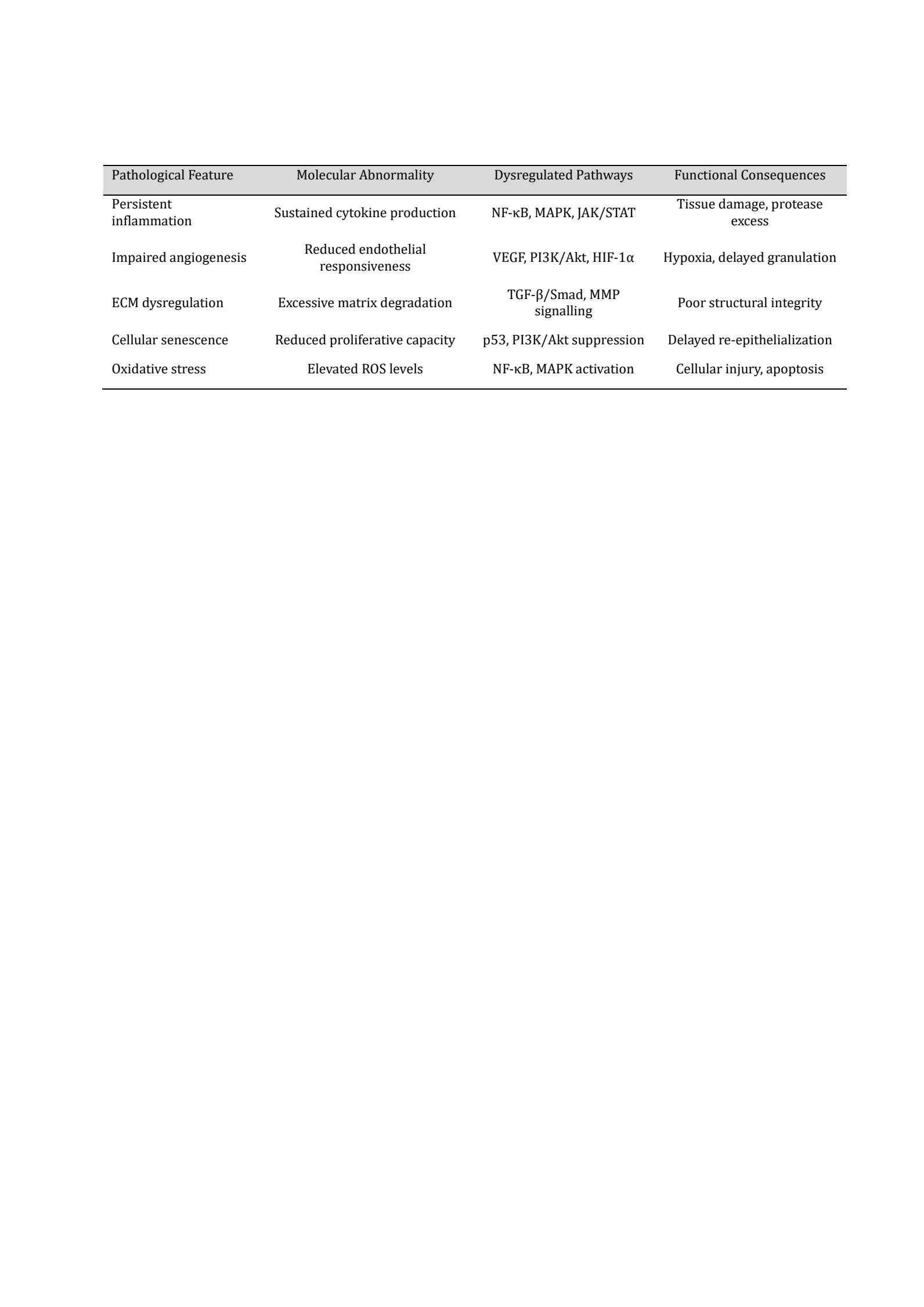
Table 3: Pathophysiological Features of Chronic Wounds and Associated Signalling Dysregulation. Chronic wounds persist due to a self-reinforcing microenvironment created by sustained inflammation, defective angiogenesis, extracellular matrix breakdown, cellular senescence, and hypoxia-induced oxidative stress, which together disrupt key regenerative signalling pathways (Figure 2)
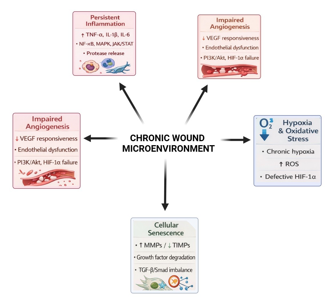
Fig. 2: Pathophysiological hallmarks of chronic wounds. This schematic is depicted as the multifactorial processes which maintain chronic wound pathology. The microenvironment of the chronic wound that is constantly formed under the influence of five interdependent pathological processes. Acute inflammation leads to persistent inflammation due to the continued presence and dominance of neutrophils and the subsequent production of high levels of pro-inflammatory cytokines (TNF-a, IL-1b, IL-6) and sustained activation of NF-kB, MAPK, and JAK/STAT signalling pathways and excessive release of proteases. Impaired angiogenesis occurs due to endothelial dysfunction and decreased sensitivity to vascular endothelial growth factor (VEGF) through dysfunctional PI3K/Akt and HIF-1a signalling and results in the deficiency of neovascularization and tissue perfusion. The dysregulation of extracellular matrix (ECM) is marked by a disproportion of the presence of matrix metalloproteinases and their inhibitors (MMPs/TIMps), an increased rate of collagen degradation, and disturbed TGF-b/Smad signalling, which leads to an unstable wound matrix. Cell-cycle arrest, p53 activation, and secretion of senescence-associated secretory phenotype (SASP) are additional mechanisms that weaken tissue regeneration through cellular senescence of fibroblasts and keratinocytes. The cellular dysfunction and signalling failure are aggravated by hypoxia and oxidative stress characterized by the chronic lack of oxygen, the overproduction of reactive oxygen species (ROS), and impaired stabilisation of HIF-1a. Together, such interrelationships strengthen a negative wound environment that does not allow the process to proceed towards successful healing.
6. Major Signalling Pathways in Chronic Wound Healing
The nine pathways described in this section are presented individually for clarity, but they are not independent entities in chronic wounds: as summarised in Section 7.3 and returned to in the Discussion, extensive crosstalk links these axes into an interconnected network, and it is this network-level dysregulation, rather than isolated pathway failure, that is proposed to underlie the persistence of chronic wounds.
6.1 TGF-b/Smad Signalling Pathway This pathway involves several complex interactions between ligand, receptor and transcription factor otherwise known as agonist-antagonist interactions. Transforming growth factor-b (TGF-b) signalling pathway is one of the most important regulators of wound healing that has pleiotropic effects on inflammation, fibroblast activation, extracellular matrix (ECM) production and tissue remodelling (76). TGF-b is a molecular switch that induces the switching between inflammation and tissue rebuilding in normal wound repair. Nevertheless, during chronic wounds, the TGF-b / Smad signalling is impaired, which leads to poor healing processes. TGF-b ligands stimulate the type II TGF-b receptors which in turn recruit type I receptors and phosphorylate them (77). The receptor complex activates receptor-mediated Smad proteins (Smad2 and Smad3) that in turn interact with Smad4 and translocate to the nucleus in a bid to mediate gene transcription. These transcription programmes regulate the synthesis of collagen, proliferation of fibroblasts and differentiation of myofibroblasts (78). It has been highlighted in the literature that it is important that this pathway be activated with precision, i.e., early activation is required to promote repair, but continued or excessive activation is pathological. In chronic wounds, TGF-b signalling is usually incapacitated regardless of the increase in ligand levels. High levels of protease activity impair TGF-b receptor and co-receptors thereby decreasing the signal transduction. Inflammatory cytokines also disrupt activation of Smads by enhancing the expression of Smad7 that inhibits phosphorylation by receptors. This leads to the inability of fibroblasts to generate structured ECM, and contraction of the wound is impaired (79). Also, TGF-b signalling is altered, which influences the immune regulation. In normal conditions, TGF-b inhibits excess inflammation by down-regulation of production of pro-inflammatory cytokines. TGF-b decreased responsiveness in chronic wounds maintains increased inflammatory signals that enhance tissue damage. However, in other situations, aberrant TGF-b stimulation can stimulate fibrosis and overgrowth instead of functional regeneration. The duality of TGF-b also points to the difficulty of therapeutically targeting this pathway (80). Mechanisms that are effective should reinstate physiological signalling harmony instead of merely augmenting or repressing the activity of pathways. The contextual regulation of TGF-b/Smad signalling is still the key to designing specific therapy to catheterize the chronic wound healing.
6.2 Wnt/b-Catenin Signalling Pathway WNT/b-catenin signalling is essential in regeneration of the epidermis, activation of stem cells and patterning of tissues during wound healing. Transient Wnt signalling by normal repair stimulates keratinocyte proliferation and migration and leads to rapid re-epithelialization. The pathway mal-regulation is intimately associated with the inability to repair epithelia in chronic wounds (81). Canonical Wnt signalling Wnt ligands that bind Frizzled receptors and LRP5/6 co-receptors prevent the destruction complex of b-catenin. The accumulated b-catenin stabilized in the cytoplasm translocates to the nucleus controlling transcription of cell proliferating, cell migrating, and cell maintenance genes. This signalling is spatially limited in the case of acute wounds to the wound edge and basal epidermal layers (82). WNT/b-catenin, in chronic wounds, is usually diminished or improperly localized. Nuclear translocation is disrupted and b-catenin is destabilized by oxidative stress and chronic inflammation. Keratinocytes are also modulated in expression of receptors by inflammatory mediators, turning them unresponsive to Wnt ligands. Consequently, keratinocyte migration is reduced and re-epithelialization is postponed (83). WNT signalling defects are further complicated by stem cell dysfunction. Hair-follicle-derived and interfollicular stem cells rely on Wnt signalling to exit quiescence and contribute to wound repair; impaired Wnt signalling in chronic wounds therefore reduces regenerative capacity by limiting stem-cell-driven epithelial repair. Prolonged or excessive Wnt activation is equally problematic, however, resulting in abnormal differentiation or fibrosis (84). This underscores the importance of the balanced Wnt signalling in the course of effective healing. The pathway therapy must be carefully timed and spatially controlled to prevent the negative effects.
6.3 PI3K/Akt/mTOR Signalling Pathway PI3K/Akt/mTOR pathway is a key regulator of cell survival, metabolism, migration and angiogenesis in wound healing. This signal transduction pathway combines all growth factor, cytokine, and extracellular matrix interaction signals into a series of cellular responses. The studies are continually pointing a similar defect with impaired PI3K/Akt signalling in chronic wounds, especially in diseased conditions of a metabolic malfunction like diabetes (85). This pathway is activated by stimulating receptor tyrosine kinases with growth factors which activates phosphoinositide 3-kinase (PI3K) and phosphatidylinositol-3, 4,5-trisphosphate is produced. This lipid second messenger recruits Akt to the plasma membrane, which is then activated and controls downstream pathways, such as mTOR. These signals together stimulate protein synthesis and cell survival coupled with cytoskeletal rearrangement (86). PI3K/Akt signalling promotes the migration of keratinocytes, fibroblast proliferation and endothelial cell survival in normal wound healing. It also increases the production of nitric oxide that activates angiogenesis. In chronic wounds, the activation of Akt by oxidative stress and inflammatory mediators leads to decreased cellular responsiveness to growth factors. This is further disturbed in hyperglycemia which activates phosphatases that inactivate Akt (87). mTOR pathology is one of the causes of maladaptive protein synthesis and metabolism in wound cells. The low activity of mTOR inhibits the production of the fibroblast matrix and cell proliferation of the keratinocytes, which halts tissue regeneration. Restoring PI3K/Akt/mTOR signalling, the wound-healing process is improved in experimental models, which makes it therapeutically relevant (88).
6.4 MAPK (ERK, JNK, p38) Signalling Pathways The mitogen-activated protein kinases (MAPK) signal transduction mechanisms play an important role in the wound healing process by mediating inflammation, cell proliferation, migration, and cell stress responses. The three branches of MAPK namely, ERK, JNK and p38, have different yet connected roles. Dysregulated MAPK signalling is one of the factors that leads to chronic inflammation and impaired repair in chronic wounds. Cell proliferation and migration are mainly linked to the ERK signalling. During normal healing, the ERK activation stimulates the growth of keratinocytes and fibroblasts in the proliferative stage. The insufficiency or transient nature of ERK signalling in chronic wounds limits regenerative ability (89-93). Stress stimuli, such as oxidative stress and inflammatory cytokines, on the other hand, trigger JNK and p38 pathways. Transient activation is helpful in immune defense whereas persistent activation is facilitated to promote apoptosis, cytokine production, and tissue damage. The non-healing wounds have a sustained JNK and p38 activation that strengthens the inflammatory signalling and prevents the tissue regeneration. The imbalance of the MAPK pathway so changes the wound environment towards catabolism as opposed to regeneration (94). Precise targeting of MAPK signalling is a potential therapeutic strategy that can be employed to restore healing balance.
6.5 NF-kB Signalling Pathway NF-kB is a key regulator of inflammatory gene expression and immune responses during wound healing. NF-kB activation in acute wounds is short and quick and is adequate to protect against pathogens. NF-kB signalling however becomes permanently active in chronic wounds, leading to pathological inflammation. NF-kB is activated by degradation of inhibitory IkB proteins and NF-kB dimers can then move to the nucleus and initiate transcription of cytokines, chemokines and adhesion molecules. Chronic wounds have high rates of TNF-a, IL-1b, and IL-6 due to the sustained NF-kB activity (95). Constant NF-kB signalling suppresses the migration of keratinocytes, activates the production of proteases, and lowers growth factor signalling. It also disrupts macrophage polarization, which does not allow switching to reparative phenotypes (96). Selective inhibition of NF-kB with little or no effect on host defence would therefore be of great interest in the context of therapeutic strategies.
6.6 JAK/STAT Signalling Pathway The Janus kinase/signal transducer and activator of transcription (JAK/STAT) pathway is an immunomodulator-mediated cytokine-based communication between immune cells and skin-resident cells. This pathway is important in controlling wound healing inflammation, proliferation of cells, and immune resolution. To promote transcription of phosphorylated and translocated nuclear STAT, cytokine binding causes JAK kinases to be activated (97). Prolonged JAK/STAT activation which is sustained by excess cytokine signalling in chronic wounds supports sustained expression of inflammatory genes. The development of chronic inflammation involves the aberrant STAT activation that interferes with the macrophage polarization and the work of fibroblasts (98). The JAK/STAT signalling would provide opportunities to adjust immune reactions and provide wound homeostasis.
6.7 The Hippo–YAP/TAZ Signalling Pathway The Hippo pathway regulates organ size, cell growth and tissue renewal by use of YAP and TAZ transcriptional co-activators. Mechanical cues and cell density during wound healing regulate YAP/TAZ activation, which facilitates YAP/TAZ migration and proliferation of keratinocytes. In chronic wounds, the activity of YAP/TAZ is impaired by impaired mechanical signalling and inflammatory stress. The decreased Hippo signalling inhibits epithelial regeneration and granulation tissue formation, which causes slow healing (99).
6.8 Notch Signalling Pathway Notch signalling regulates cell differentiation, epidermal homeostasis, and angiogenesis. Subsequent interactions between ligands and receptors between adjacent cells activate it, and the differentiation-related genes are transcriptionally regulated. In chronic wounds, the impaired keratinocyte differentiation and vascular stability are caused by the altered Notch signalling. Unregulated Notch signalling also helps in maladjusted epithelial stratification and dysfunctional angiogenesis (100).
6.9 Mediating Hypoxia Signalling through HIF-1a One of the major regulators of adaptation to low oxygen in cells is hypoxia-inducible factor-1a (HIF-1a). HIF-1a increases angiogenesis, glycolytic metabolism, and cell survival in acute wounds. In chronic wounds, the HIF-1a signalling, however, is often defective, as a consequence of oxidative stress and metabolic dysfunction. Dysfunctional HIF-1a signalling limits VEGF induction and the angiogenic response, sustaining the ischemia. Rescue of hypoxia-sensitive signalling is a strong potential line of therapeutic intervention in chronic wound healing (101). The major intracellular signalling pathways implicated in chronic wound healing, their biological roles, patterns of dysregulation, and therapeutic implications are summarized in Table 4.
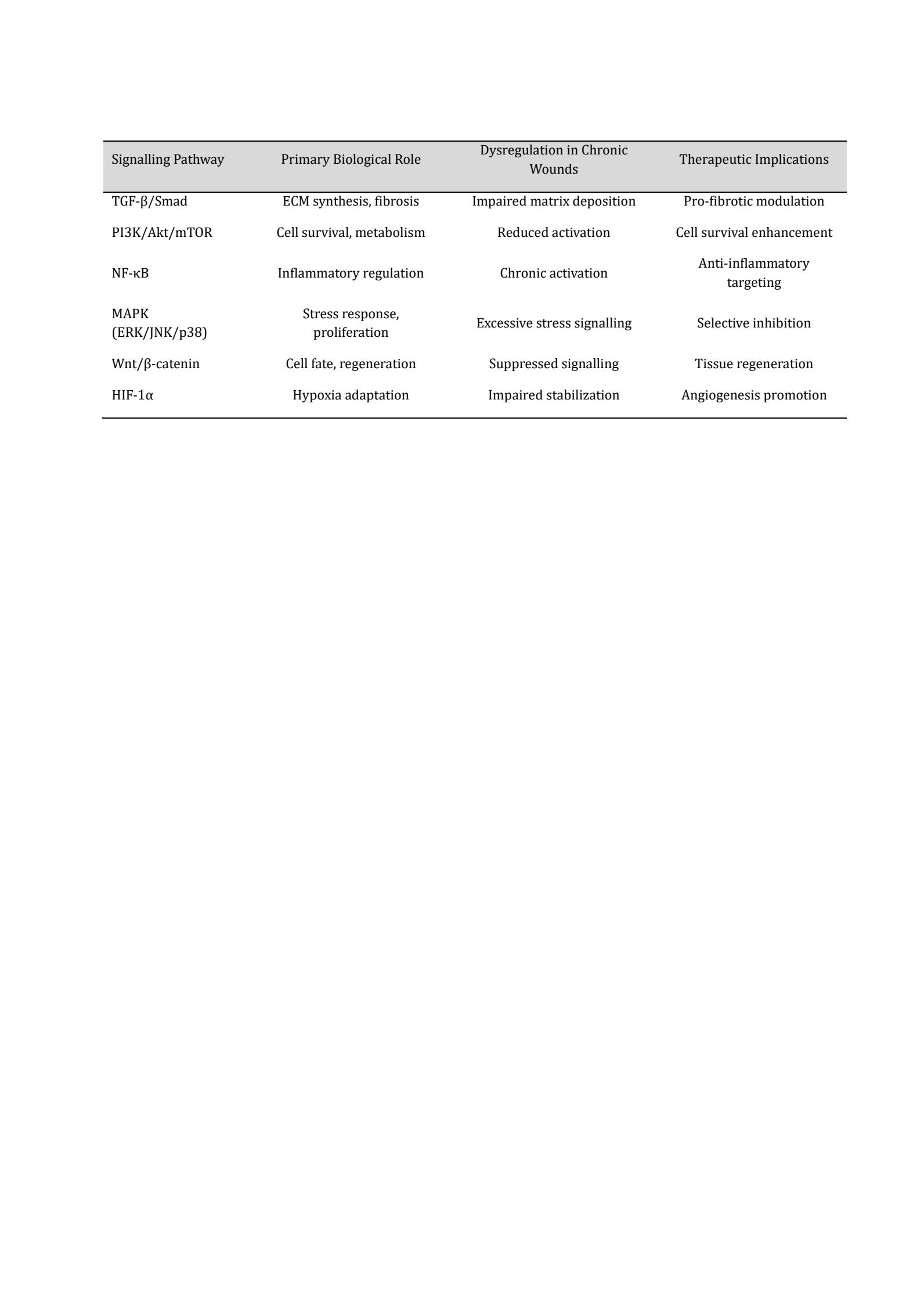
Table 4: Key Signalling Pathways Implicated in Chronic Wound Healing
7. Inflammatory and Immune Signalling in Chronic Wounds
7.1 The role of Macrophage Polarization (M1/M2) Macrophages are the primary immune controllers of wound healing, acting as dynamic regulators of both inflammatory and reparative cues. In normal wound repair, macrophages are highly plastic, shifting from a pro-inflammatory phenotype in early injury to a reparative phenotype during tissue regeneration. This phenotypic transition fails to occur successfully in chronic wounds (102). At the initial inflammatory stage, the macrophages shift towards a classical activated/pro-inflammatory phenotype, which involves the generation of cytokines e.g., TNF-a, IL-1b and IL-6. These mediators increase pathogen clearance and attract more immune cells. NF-kB, JAK/STAT and p38 MAPK are some signalling pathways that drive this phenotype (103). This activation in acute wounds is limited and short-lived. The macrophages in healing normally transition to an alternatively activated reparative phenotype as the healing progresses. VEGF, PDGF and TGF-b are the growth factors released by the reparative macrophages, which encourage angiogenesis, fibroblast activation, and deposition of the extra-cellular matrix. The process of this transition is facilitated by cytokine exposure rearrangements, metabolic re-organization and intra-cellular signalling readjustments (61). In chronic wounds, macrophages tend to remain trapped in the pro-inflammatory phase. Constant exposure to inflammatory cytokines, oxidative stress, and metabolic dysregulation inhibits phenotypic switching. Hyperglycaemia, ischaemia, and microbial burden alter macrophage signal transduction, sustaining NF-κB and STAT activation while repressing reparative signalling pathways. This imbalance has severe effects on wound healing (104). Pro-inflammatory macrophages also secrete proteases and reactive oxygen species that break down extracellular matrix components and growth factors. Meanwhile, the lack of reparative mediators restrains fibroblast proliferation, angiogenesis, and epithelial regeneration. The wound environment thereby becomes self-perpetuating, as inflammation intensifies tissue destruction and blocks healing (105). One of the opportunities of therapy is the restoration of macrophage plasticity. Instead of the general immunosuppression, targeted control of the macrophage signal transduction can provide a solution to restore the inflammatory-reparative balance that is required to resolve a wound.
7.2 Chronic Inflammation Cytokines and Chemokines The major communication system in wound healing by the immune system is made up of cytokines and chemokines. These soluble mediators are generated in a carefully controlled time program which is effective defence followed by resolution in acute wounds. This regulatory system fails in the case of chronic wounds, which leads to prolonged cytokine signalling and perpetuation of inflammation (106). TNF- a, IL-1b and IL-6 are known pro-inflammatory cytokines which are consistently higher in chronic wounds. These cytokines promote intracellular signalling pathways such as NF-kB, MAPKs, which promote transcription of inflammatory genes and proteolytic enzymes. The persistent exposure to these mediators has been shown to inhibit the migration of keratinocytes, inhibit the activity of fibroblasts and disrupt the extracellular matrix constituents. The chemokines also enhance the inflammation by constantly bringing the immune cells to the wound (107). The continuous chemokine gradients prevent the elimination of immune response and ensure a high cellular density of the wound bed. This overload of immune response enhances oxidative stress and protease action thus worsening tissue damage. The anti-inflammatory cytokines normally counter the effect of inflammatory signalling are inadequate or ineffective in chronic wounds. There is also lowered sensitivity of regulatory cytokines which worsens resolution (107). Persistent inflammation also alters the expression of cytokine receptors and downstream signalling responses, causing abnormal physiological responses despite the presence of growth factors. Cytokine signalling in chronic wounds does not act in isolation: cytokines are closely connected to growth factor pathways, mechanical signals, and metabolic signals. Persistent inflammatory cytokines effectively disrupt regenerative signalling, suppressing PI3K/Akt and Wnt signalling and thereby impairing tissue repair (17). The cytokine network dysregulation is important in the understanding and development of specific therapies that inhibit pathological inflammation without affecting the vital immune defences.
7.3 Cross-Talking between Immune-cells and Skin Cells The communication between the immune cells and the skin-resident cells, such as keratinocytes, fibroblasts, and endothelial cells, is constant to ensure the successful wound healing. This two-way crosstalk aligns immune defence and tissue regeneration. In chronic wounds, the broken interaction among these cells groups plays a large role in poor healing. The immune cells control the behaviour of skin cells via cytokine and growth factor signalling (108). Macrophage-mediators control the proliferation of fibroblasts, their matrix as well as angiogenesis. Keratinocytes also act on immune cues by changing migration, differentiation, and restoring the barrier. In normal healing, these interactions are stage-specific adaptive interactions (109). This communication network is dominated by immune-derived inflammatory signals in chronic wounds. Chronic exposure to cytokines causes changes in gene expression of the keratinocytes reducing their migratory ability and aberrant differentiation. Chronic inflammatory stimulated fibroblasts have decreased matrix production and could be senescent. On the other hand, dysfunctional skin cells also affect immune behaviour. Injured keratinocytes secrete danger-associated molecular patterns which sustain immune activation. Damaged fibroblasts are not able to give structural and biochemical signals of immune resolution. Endothelial dysfunction restricts the immune cells traffic and angiogenesis(5, 110). The interrupted crosstalk provides a feedback mechanism whereby immune dysfunction and tissue failure mutually reinforces each other. To correct such a breakdown in communication, it would be necessary to induce specific changes in signalling pathways at the immune-skin interface instead of specifically changing a single cell type.
8. Angiogenic and Vasculogenic Signalling Targets
8.1 VEGF-Dependent Pathways The main mediator of angiogenesis in wound healing is vascular endothelial growth factor (VEGF) signalling, which is critical in the process of restoring tissue perfusion following the wound. Under normal wound repair conditions, hypoxia and inflammatory signals trigger VEGF expression that subsequently triggers endothelial cell proliferation, migration, and survival. The VEGF signalling is required to mediate new vessel formation, as well as vascular permeability and extracellular matrix remodelling (111). VEGF has effects which are mainly mediated by binding to VEGF receptor-2 on endothelial cells. Receptor autophosphorylation and activation of downstream signalling, such as PI3K/Akt and MAPK/ERK signalling is stimulated by ligand-receptor interaction. These signalling pathways enhance survival of endothelial cells, reorganization of cytoskeleton as well as directional migration of the cells towards angiogenic factors. VEGF signalling in acute wounds is highly localized to regions of the wound bed hypoxia with a decrease in hypoxia as the wound bed is vascularized (112). VEGF signalling is often dysfunctional in chronic wounds even in the presence of ongoing hypoxia. Oxidative stress and inflammatory mediators inhibit VEGF receptor activation and downstream signal transduction. Endothelial cells show reduced responsiveness to VEGF, resulting in ineffective or poorly organised vessel formation, while excessive protease activity degrades VEGF and its receptors, further reducing signalling efficiency (113). Disrupted VEGF signalling is one of the factors contributing to insufficient oxygen and nutrient supply, which in turn sustains hypoxia and metabolic stress in the wound. This forms a vicious cycle in which hypoxia fails to induce effective angiogenesis, increasing wound chronicity. Simply increasing ligand availability is therefore less important than therapeutic measures that restore VEGF pathway responsiveness (114).
8.2 PDGF and FGF Signalling Platelet-derived growth factor (PDGF) and fibroblast growth factor (FGF) signalling pathways are complementary, together stimulating fibroblast recruitment, matrix deposition, and angiogenesis in wound healing. Specifically, these pathways promote the formation and stabilisation of new blood vessels by supporting granulation tissue formation. Platelets are the earliest source of PDGF release, with macrophages and endothelial cells subsequently contributing further PDGF as healing progresses. PDGF binding to its receptor triggers intracellular cascades, including PI3K/Akt and MAPK, that promote fibroblast proliferation and migration into the wound bed. PDGF-mediated pericyte recruitment also supports vessel maturation and stability (115). FGFs, specifically FGF-2s promote proliferation of endothelial cells as well as promoting angiogenic sprouting. FGF signalling promotes migration of the keratinocytes and fibroblast activity in a synergistic manner with VEGF. In healthy tissue repair, the PDGF and FGF signalling have been closely controlled to achieve balanced tissue development. PDGF and FGF signalling is usually suppressed in chronic wounds. The pathway is inhibited by receptor downregulation, ligand proteolytic degradation, and intracellular signalling malfunction (116). The compromised responsiveness instead of the lack of the ligand is a significant impediment, which is why the application of growth factor therapies to clinical environments has been achieved only with limited success. Re-initiation of PDGF and FGF signalling activity is one of the major therapeutic intervention goals.
8.3 Endothelial Dysfunction in Chronic Wounds Endothelial dysfunction has become a focal pathological phenomenon in the pathogenesis and maintenance of chronic wounds. The vascular endothelium is central to sustaining tissue homeostasis by modulating vascular tone, vascular permeability, leukocyte trafficking, angiogenesis, and coagulation. A normal endothelium is important for wound healing, promoting blood flow, oxygen and nutrient supply to the wound, and the development of new microvasculature (117). A decrease in endothelial integrity and function disturbs the well-orchestrated wound repair process, putting wounds at risk of chronicity. Endothelial cells in chronic wounds display structural damage and functional impairment associated with low nitric oxide (NO) bioavailability, oxidative stress, and a persistent pro-inflammatory phenotype (118). NO is one of the key endothelial-mediators that facilitate vasodilation, platelet aggregation inhibition and angiogenesis. The damage of endothelium causes decreased production of NO, which causes vasoconstriction, a decrease in perfusion, and tissue hypoxia. Hypoxia is only temporarily useful in wound healing in acute wounds but is harmful when sustained, as in chronic wound healing, where the migration of keratinocytes, fibroblast action and collagen formation are inhibited by hypoxia (119). Another characteristic of chronic wounds is persistent inflammation, which is strongly interconnected with endothelial dysfunction. Damaged or stimulated endothelial cells express adhesion molecules and release pro-inflammatory cytokines (tumour necrosis factor-α) and chemokines that sustain leukocyte recruitment. Although immune cell infiltration is required during the early inflammatory stage of wound healing, its continued presence results in excessive protease activity, extracellular matrix (ECM) breakdown, and additional endothelial injury (120). This forms a vicious, self-perpetuating cycle in which inflammation and endothelial dysfunction reinforce each other, preventing the wound from progressing to the proliferation and remodelling stages of healing. Endothelial dysfunction also has a dramatic impact on angiogenesis, an essential process in granulation tissue formation. Dysregulated angiogenic mediators in chronic wounds include vascular endothelial growth factor (VEGF) and angiopoietins. Although VEGF levels can be high, endothelial responsiveness to these signals is often inadequate, manifesting as endothelial cell apoptosis, poor migration, and poor tube formation (121). There is a further contribution of immature and leaky neo vessels by an imbalance between angiopoietin-1, which maintains the stability of blood vessels, and angiopoietin-2, which induces vascular destabilization. Consequently, the granulation tissue is never well vascularized and does not have sufficient functional capacity to aid wound closure (122). The endothelial dysfunction of chronic wounds also overlaps with the transformation of growth factor-b (TGF-b) signalling. TGF-b is important in the regulation of inflammation, fibroblast activation, myofibroblast differentiation and ECM deposition. TGF-b expression and signalling changes have been linked to endothelial injury resulting in inadequate myofibroblast availability and retarded collagen expression (123). These alterations impair tensile strength of the wounds and retard re-epithelialization which is seen in non-healing wounds. Together endothelial dysfunction leads to the development of chronic wound pathology by reducing perfusion, perpetuating inflammation, interfering with angiogenesis and modulating growth factor signalling. The evidence indicates that chronic wounds are not only a focal defect of the skin but the expression of vascular pathology. The therapeutic approach focused on endothelial functional recovery, including but not limited to increasing the bioavailability of NO, releasing oxidative stress, balancing inflammatory reactions, and restoring angiogenic and TGF-b signalling, has great potential. Attacking the endothelium can thus be a convergent and useful intervention to interrupt the pattern of lower healing and lead to proper healing of chronic wounds (124).
9. Novel Molecular Targets and regulating factors
9.1 MicroRNA and Long Non-Coding RNA The microRNAs (miRNAs), long non-coding RNAs (lncRNAs) have become the key players of post-transcriptional regulation of gene expression during the wound healing process. In contrast to protein-coding genes, these non-coding RNAs, control cellular responses by regulating mRNAs stability, translation and chromatin organization. Non-coding RNAs are dysregulated, which plays a role in the disruption of signalling in the chronic wounds (125). The miRNAs are small RNA molecules which inhibit gene-transcription through binding with complementary sequences on target mRNAs (126). Certain miRNAs that control inflammation, angiogenesis, and keratinocyte migration are activated in normal wound healing by acting on important signalling components. As an example, some miRNAs tone down overproduction of inflammatory signalling through NF-kB activation, whereas others stimulate angiogenesis through stabilisation of VEGF pathways (127). There is a significant change in the expression profile of miRNA in chronic wounds. Pro-inflammatory miRNAs are usually increased which helps in maintaining the production of cytokines and the recruitment of immune cells. On the other hand, angiogenic and epithelial regenerative miRNAs are suppressed. It is an imbalance that alters the intracellular signalling networks and abates the cell responsiveness to growth factors (128). Long non-coding RNAs can have an action in different ways, such as scaffolding protein complexes, controlling chromatin accessibility and controlling the activity of transcription factors. The lncRNAs affect the fibroblast activation, endothelial cell activity, and the polarization of macrophages. In chronic wounds, the expression of lncRNA is aberrant and this leads to prolonged inflammation and impaired remodelling of the matrix of the tissue. A possible approach to reestablish signalling equilibrium is to target non-coding RNAs (129). They are promising therapeutic targets because of their capability to control various genes at once, but delivery and specificity issues still exist.
9.2 Signalling by Exosomes and Extracellular Vesicles Exosomes and other extracellular vesicles (EVs) are becoming important mediators of intercellular communication during wound healing. These vesicles are nanoscale and carry proteins, lipids and nucleic acids across cell boundaries which allows a coordinated response throughout the wound microenvironment. The EV-mediated signalling is a key to regulate inflammation, angiogenesis and tissue regeneration. Under healthy wound repair, EVs secreted by immune cells, fibroblasts, keratinocytes, and stem cells transmit regulatory signals, which enhance healing (130). The exosomes of stem cells, especially the ones of the derivative fabric, transmit miRNAs and growth factor-related proteins that stimulate angiogenesis and fibroblast activities. Target cells have intracellular pathways (PI3K/Akt and MAPK) that are activated by these vesicles to aid survival and migration (131). EV signalling is dysregulated in chronic wounds. The EVs discharged by inflammatory cells contain pro-inflammatory mediators, which supplement the immune activation. Simultaneously, regenerative EV signals are reduced because of stem cell dysfunction and cellular communication. This disproportion leads towards inaccurate signalling coordination in the bed of the wound (132). Exosome-based strategies represent a promising therapeutic opportunity: compared to cell-based therapies, exosomes are less immunogenic and simpler to standardise. This evidence remains preclinical: in animal chronic-wound models, administration of regenerative EVs has been reported to restore angiogenic and anti-inflammatory signalling, but human clinical data are not yet available. Nevertheless, there are still issues with the isolation, characterization and delivery of EVs (133). This will be critical in understanding the EV cargo composition and targeting mechanisms and translating the same to clinical use.
9.3 Epigenetic Control of Wound Healing Mechanisms Epigenetic regulation is very important in regulating the expression of genes in wound healing without modifying the basic DNA sequence. Cellular responsiveness to signalling cues is regulated by epigenetic mechanisms such as DNA methylation, histone modifications and chromatin remodelling. The epigenetic disturbance is one of the points that promote the maintenance of pathological signalling of chronic wounds (134). During the normal wound healing, epigenetic events enable cells to change their gene expression programs at a rapid rate in response to injury. As an illustration, histone acetylation increases inflammatory and proliferation genes accessibility at the early phases and subsequently suppresses these genes in later stages to promote resolution and remodelling (135). Persistent inflammatory signalling causes durable changes in epigenetics in chronic wounds, which entrap cells in maladaptive positions. There are also changes in the methylation of fibroblasts and immune cells which inhibit regenerative genes and up regulate inflammatory genes. Such epigenetic changes decrease the plasticity of cells and minimize an ability to respond to growth factors (136). Hyperglycemia, hypoxia, and oxidative stress are other elements of the environment that have an impact on the epigenetic control. The chromatin-modifying enzyme activity is modified by the metabolic stress, which strengthens chronic wound pathology (137). Epigenetic therapies hold a new avenue of intervention. These techniques can also reestablish normal signalling dynamics by reprogramming gene expression profiles. Specificity and safety are however key considerations.
10. Therapeutic Interventions of Signalling Pathways
10.1 Small-Molecule Inhibitors and Activators Small-molecule therapeutics: one of the most flexible approaches to regulating dysregulated signalling pathways in chronic wound healing. The agents are geared to inhibit or activate directly the major intracellular signalling components in a selective manner, which will restore the balance between the inflammatory and regenerative processes. The small molecules have the benefit of controlled dosing, tissue penetration, and ability to control intracellular targets that biologics do not have access to (10). The activation of inflammatory pathways like NF-kB and stress-related MAPKs persists in chronic wounds, and it increases the degradation of the tissue and the impairment of repair. Small-molecule inhibitors of these pathways have reduced excessive cytokine release and protease activity in preclinical (cell culture and animal) models; these agents reduce the pathological inflammation, which makes the microenvironment more favourable to tissue regeneration (138). On the other hand, small molecule agonists of regenerative signalling seek to improve signalling that is impaired in chronic wounds. Compounds that increase PI3K/Akt/HIF-1α signalling have been shown to promote cell survival, angiogenesis, and metabolic adaptation (128). Restoring downstream signalling sensitivity is often more effective than simply increasing growth factor levels. Small-molecule therapies have specificity issues, and systemic effects are a challenge to their clinical potential. The signalling pathways involved in wound healing also regulate key physiological processes in other tissues. (139) Thereby, localized delivery and formulations are essential to reduce off-target effects and the therapeutic benefit as much as possible.
10.2 Growth Factors and Biologics Biologic agents and growth factors have long been considered as therapeutic modulators of chronic wound healing because of their core role in regulating cell behaviour. Growth factors such as PDGF, VEGF and FGF directly stimulate cell proliferation, migration, and angiogenesis, but clinical responses to growth factor therapy have been variable. Becaplermin (recombinant PDGF-BB) remains the only growth-factor formulation with regulatory approval for chronic wound care (diabetic foot ulcers); most other growth-factor and biologic approaches remain at the preclinical or early-phase clinical stage (Table 5). The first limitation is disrupted growth factor signalling in chronic wounds. Cellular responsiveness is inhibited by receptor downregulation, protease-mediated degradation, and intracellular signalling defects (2). This leads to the fact that a mere rise in ligand concentration does not usually regain healing. Biologic treatment of inflammatory mediators has also received investigation. Neutralization of pro-inflammatory cytokines with antibodies is expected to eliminate pathological inflammatory responses and induce resolution. Although these therapies have been shown to be effective in the laboratory, they have proven difficult to apply to wound care because of the risk of infection and the weakening of the immune system. The combination strategies, meaning combining growth factors with signalling modulators or biomaterials, could be more effective, defending ligands or promoting receptor interactions (140).
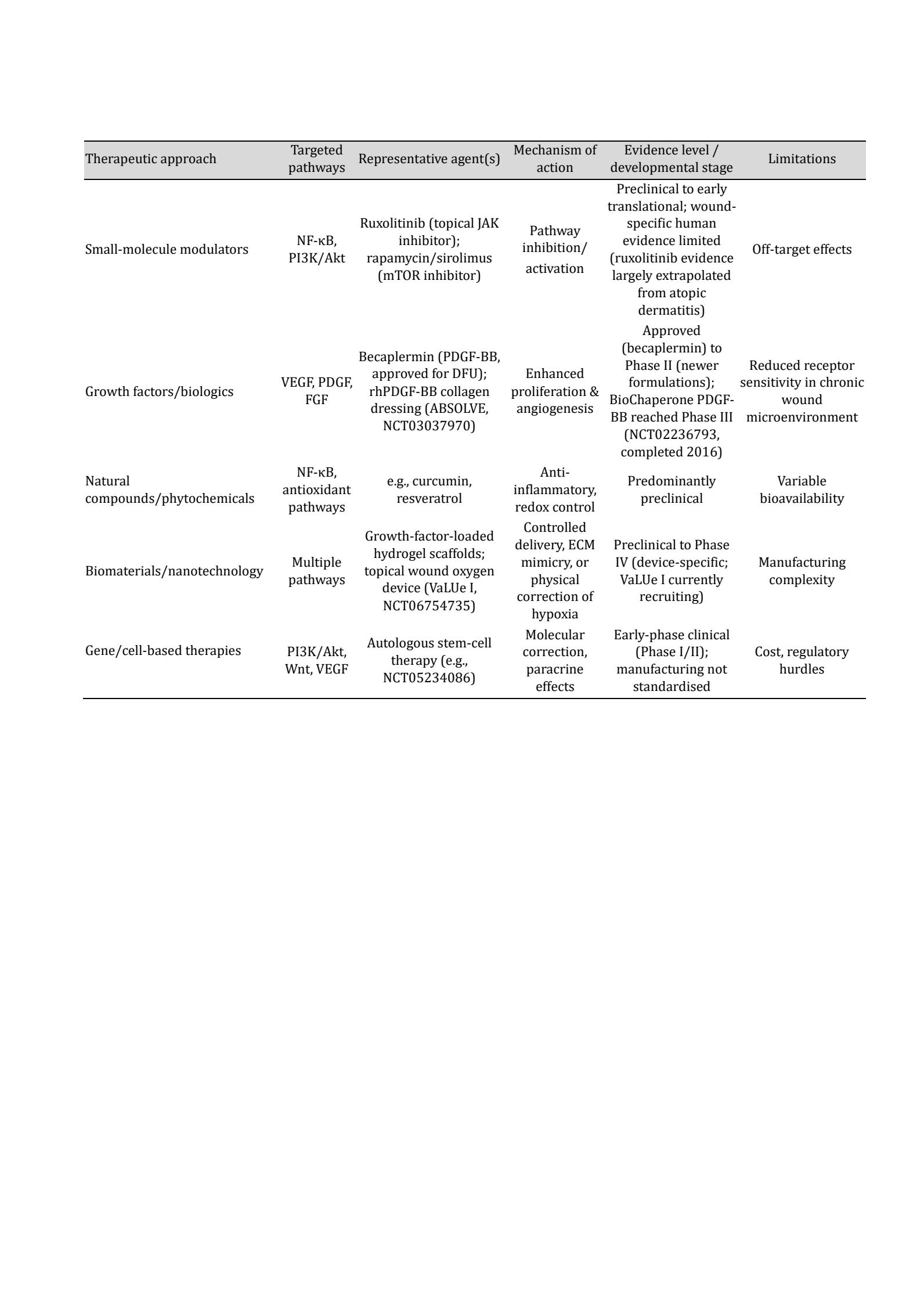
Table 5: Therapeutic Strategies Targeting Signalling Pathways in Chronic Wound Healing, with Representative Agents and Evidence Level
10.3 Natural Compounds and Phytochemicals Because of the multi-targeted nature of their biological actions, natural compounds and phytochemicals have become more and more popular as adjunctive agents in chronic wound care. Natural agents have anti-inflammatory, antioxidant, and pro-angiogenic actions, which means that they can regulate multiple signalling pathways at a time (141). The natural and plant-derived compounds have the ability to inhibit NF-kB, decrease oxidative stress, and stimulate growth factor signalling. These compounds have a wider regulatory effect, in contrast to highly specific synthetic agents, and can be beneficial in the complicated pathology like chronic wounds. Nevertheless, inconsistency in bioavailability, strength and formulation is a major effect (142). Evidence for natural compounds in chronic wound healing remains predominantly preclinical (cell culture and animal models); controlled clinical trials in chronic human wounds are limited, and standardisation and controlled delivery systems will be needed to provide reproducible therapeutic results.
10.4 Biomaterials and Nanotechnology Solutions Nanotechnology-based approaches and biomaterials are an emerging field of interest in chronic wound care. These strategies are meant to offer structural assistance, shield therapeutic agents and allow regulated discharging of signalling modulators. The biomaterials do not have to be passive wound dressings but can act to have an active response to cellular signalling. The more complex scaffolds and hydrogels are created to replicate the extra-cellular matrix, including biochemical and mechanical signals that enhance cell attachment and movement (143). Nanoparticles can be designed to be able to present growth factors, small molecules, or nucleic acids to the desired cells, increasing the precision of the treatment. Biomaterials provide a solution to centrally approaches to wound therapy combining physical support with a molecular signalling modulation to deal with several facets of chronic wound pathology (144). A recently described emerging technology, plasma-activated gas, illustrates this biomaterial-adjacent direction: non-thermal plasma-generated reactive oxygen and nitrogen species have been reported to activate VEGFR-ERK1/2 and TGF-β/Smad signalling and to rebalance MMP/TIMP activity in preclinical and early clinical wound-healing studies, in addition to broad-spectrum antimicrobial and biofilm-disrupting effects (156).
10.5 Gene and Cell-Based Therapies The most developed and personalized methods of addressing the signalling pathways in the chronic wound healing include gene and cell-based therapies. These approaches seek to remedy an underlying defect in the molecules by offering functional genes or regenerative cells so that they are directly delivered to the wound. Gene therapy methods aim at increasing the expression of growth factors or signalling regulators which are lacking in intractable wounds. Cell-based therapies, especially stem or progenitor cells, are meant to reestablish regeneration both by direct tissue supply and by paracrine signalling (9, 145). Although such methods demonstrate considerable potential in preclinical and early-phase clinical studies (Table 5), most gene- and cell-based approaches for chronic wounds remain in Phase I/II testing, with safety, delivery, and manufacturing standardisation still to be established before broader clinical adoption. Further refinement and incorporation into biomaterial platforms can help increase their clinical applications. An overview of current and emerging therapeutic strategies targeting dysregulated signalling pathways in chronic wounds, including their molecular targets, mechanisms of action, and limitations, is presented in Table 5.
11. Preclinical and Clinical Evidence
11.1 In vitro and Animal Model Tests. Preclinical studies have contributed significantly to the understanding of the chronic wound pathology pathophysiology and the confirmation of signalling pathways as therapeutic targets. In vitro experiments with keratinocytes, fibroblasts, endothelial cells and immune cells have shown that chronic wounds are defined by persistent inflammatory signalling, loss of growth factor responsiveness, and membrane cellular metabolism. They have been especially helpful in breaking down pathway-specific effects, e.g. the contribution of NF-kB to cytokine production or PI3K/Akt malfunction to compromised cell survival and migration (146). The animal models, such as diabetic mice, ischemic wound models, and aged animals, are more integrative models of chronic wound healing, and they mimic some of the important clinical characteristics of chronic wounds, including slowed re-epithelialization, impaired angiogenesis, and excessive extracellular matrix degradation. The role of these models in wound healing has been proven by manipulation of signalling pathways either in genetic modification or pharmacological intervention, where restoration of pathway balance results in wound healing accelerated by a large margin (147). For example, HIF-1α signalling can be activated to enhance angiogenesis and metabolic adaptation during hypoxia, while reducing inflammatory load by inhibiting MAPK or NF-κB overactivation. These findings point to a broader pattern: the effects of any single pathway are often incomplete on their own, supporting the view that chronic wounds reflect network-level dysregulation rather than isolated molecular defects (148). Preclinical models have flaws, despite their usefulness. Animal and human skin structures, immune responses and wound contraction differ making it difficult to conduct direct translation. However, they are the studies that present important evidence of concept (proof-of-concept) in support of signalling-based therapeutic strategies.
11.2 Signalling Pathway Clinical Trials currently in progress Clinical translation of pathway-targeted therapies has been considerably slower than preclinical discovery, and only a minority of completed or ongoing trials target defined signalling intermediates directly rather than downstream tissue effects. Among growth-factor-receptor-targeted approaches, PDGF-BB-based therapies have reached the most advanced stage of clinical testing: a Phase III trial of BioChaperone PDGF-BB in chronic diabetic foot ulcers (ClinicalTrials.gov identifier NCT02236793) was completed in 2016, whereas a Phase II trial of a collagen-bound rhPDGF-BB formulation (ABSOLVE, NCT03037970) illustrates the attrition that characterises this translational pipeline, its status remaining unreported after completion of enrolment. Beyond PDGF signalling, a trial targeting the prostacyclin (PGI2) receptor pathway in diabetic foot ulcers (NCT05099367) represents an attempt to modulate cAMP–protein kinase A signalling directly, distinguishing it from growth-factor-replacement strategies. In venous leg ulcers, a Phase IV trial of cyclical topical wound oxygen therapy (VaLUe I, NCT06754735), currently recruiting, addresses the hypoxic component of the chronic wound microenvironment relevant to HIF-1α pathway dysfunction, although the intervention itself is physical rather than molecularly targeted. Cell-based and paracrine-signalling approaches, including autologous stem-cell therapies, continue to be evaluated in early-phase studies (e.g., NCT05234086) but remain limited by variable manufacturing standards and inconsistent outcome reporting. Collectively, the clinical trial landscape for signalling-pathway-targeted wound therapies remains dominated by growth-factor and device-based interventions; direct modulation of intracellular pathways such as PI3K/Akt, Wnt/β-catenin, or JAK/STAT has yet to progress beyond preclinical and early translational study in the specific context of chronic wound healing.
11.3 Limitations of Current Therapies Nevertheless, there is still a lot to be desired in terms of limitations of existing therapies of chronic wounds. One point of concern is that most of the treatments are downstream focused as opposed to upstream focused on the molecular drivers. Consequently, interventions can produce a temporary enhancement of wound appearance without restoring the signalling dysfunction (151). Patient heterogeneity is another limitation. Chronic wounds develop due to varied etiology with diabetes being one of them as well as vascular disease, pressure injuries, and aging, with each bearing their own unique molecular signature (152). Stereotyping methods of treatment do not consider this difference, which leads to uneven clinical results. More so, the chronic wound microenvironment, which is hypoxic, oxidative, protease-rich and with a microbial burden, may inactivate therapeutic agents or suppress cellular responsiveness (153). These problems highlight why both biologically-focused and context-sensitive therapies are necessary.
12. Problems and Future Outlook
12.1 Translational Barriers The process of translating signalling-targeted therapies between the bench and the bedside is still problematic. A significant obstacle is the complexity of signalling networks, which are characterised by large crosstalk and redundancy. Regulation of one pathway can result in an unwanted stimulation of compensatory responses, which restrain therapeutic effectiveness (154). Safety concerns have also been raised around manipulating cell proliferation and survival pathways, particularly for long-term or systemic therapy; local delivery and controlled modulation are important for reducing such risks. Practical barriers, including manufacturing, regulatory approval, and cost-effectiveness, present additional challenges for advanced biologics and gene-based therapies.
12.2 Customized and Accurate Medicine Precision medicine is a potential framework to nurture heterogeneity in chronic wound patients. With the combination of clinical parameters and molecular profiling, there is possibility of stratification of patients, on dominant signalling defects, and subsequently customise therapies accordingly. The biomarkers of inflammatory status, angiogenic capacity, or cellular senescence may be used to select therapies (155). Individualized strategies can enhance effectiveness and decrease the unjustified exposure to ineffective treatments.
12.3 Future Perspectives of Targeted Wound Therapy It can be expected that future research will be dedicated to combination therapies that can enable simultaneous modification of several signalling pathways. Combining biomaterials with molecular therapeutics, creating intelligent delivery methods and tapping into physiological repair processes, like exosome-mediated signalling, are among the main areas of emerging focus. Future developments in systems biology and computational modelling can improve the knowledge of signalling network dynamics and allow multi-target interventions to be rationally designed.
Conclusion
Chronic wounds are creating an increasing clinical problem of complex cellular signalling pathway dysregulation. Contrary to acute wounds, chronic wounds do not undergo sequential stages of healing because of the presence of chronic inflammation, impaired angiogenesis, defective remodelling of the extracellular matrix, and dysfunctional cells. The mounting body of preclinical and clinical research indicates that these pathological characteristics arise substantially from aberrant signalling, although chronic wounds remain multifactorial in origin, with vascular, metabolic, immunological, and microbial factors also contributing to impaired repair. Attacking critical pathways such as TGF-b/Smad, PI3K/Akt/mTOR, NF-kB, MAPK, Wnt/b-catenin, and hypoxia-responsive signalling would be a sensible approach to re-establishing the ability to heal. Nonetheless, when it comes to translation, an integrated approach must be used that takes into account the pathway crosstalk, patient heterogeneity and the hostile wound microenvironment. Improved chronic wound management in the future will rely on more accurate drug delivery, improved biomaterials, and systems-based knowledge of wound biology. Signalling-based interventions hold considerable promise for chronic wound care, although current clinical evidence for pathway-targeted therapies remains more limited than preclinical findings would suggest, and realising this potential will require translational studies that adequately capture the multifactorial nature of chronic wounds.
Acknowledgements
This research was supported by the Research, Development, and Innovation Authority (RDIA), Riyadh, Saudi Arabia, through the Reactivating Translational Medicine Lab initiative, grant number 13262-Tabuk-2023-UT-R-3-1-HW-.
Author Contributions: Conceptualization, R.M. and H.K.; methodology, R.M., W.L. and J.B.; software, M.M.J. and M.A.Alt.; validation, R.M.A., F.J.T. and M.S.M.; formal analysis, H.N.N., R.I.A. and M.A.Alan.; investigation, N.A.A. and F.H.A.; resources, A.A.B. and A.A.W.; data curation, W.L. and J.B.; writing—original draft preparation, R.M., W.L., J.B. and M.M.J.; writing—review and editing, all authors; visualization, R.M.A. and F.J.T.; supervision, R.M. and H.K.; project administration, R.M.; funding acquisition, R.M. All authors have read and agreed to the published version of the manuscript.
Data Availability Statement No new data were created or analysed in this study, as this is a narrative literature review. Data sharing is not applicable to this article.
Disclosure Statement
The authors declare no Disclosure Statement.
References
- Tottoli EM, Dorati R, Genta I, Chiesa E, Pisani S, Conti B. Skin Wound Healing Process and New Emerging Technologies for Skin Wound Care and Regeneration. Pharmaceutics. 2020;12(8). https://doi.org/10.3390/pharmaceutics12080735
- Eming SA, Martin P, Tomic-Canic M. Wound repair and regeneration: mechanisms, signaling, and translation. Sci Transl Med. 2014;6(265):265sr6. https://doi.org/10.1126/scitranslmed.3009337
- Diller RB, Tabor AJ. The Role of the Extracellular Matrix (ECM) in Wound Healing: A Review. Biomimetics (Basel). 2022;7(3). https://doi.org/10.3390/biomimetics7030087
- Liu Z, Fang Y. Wound healing and signaling pathways. Open Life Sci. 2025;20(1):20251166. https://doi.org/10.1515/biol-2025-1166
- Riaz M, Iqbal MZ, Klar AS, Biedermann T. Immunomodulatory Mechanisms of Chronic Wound Healing: Translational and Clinical Relevance. MedComm (2020). 2025;6(11):e70378. https://doi.org/10.1002/mco2.70378
- Jonidi Shariatzadeh F, Currie S, Logsetty S, Spiwak R, Liu S. Enhancing wound healing and minimizing scarring: A comprehensive review of nanofiber technology in wound dressings. Progress in Materials Science. 2025;147:101350. https://doi.org/10.1016/j.pmatsci.2024.101350
- Wang Z, Zeng X, Feng W, Lu Y, Wei P. Reframing chronic wound therapy: from growth factor delivery to regenerative immuno-engineering. Front Immunol. 2025;16:1683591. https://doi.org/10.3389/fimmu.2025.1683591
- Gumede DB, Abrahamse H, Houreld NN. Targeting Wnt/beta-catenin signaling and its interplay with TGF-beta and Notch signaling pathways for the treatment of chronic wounds. Cell Commun Signal. 2024;22(1):244. https://doi.org/10.1186/s12964-024-01623-9
- Kolimi P, Narala S, Nyavanandi D, Youssef AAA, Dudhipala N. Innovative Treatment Strategies to Accelerate Wound Healing: Trajectory and Recent Advancements. Cells. 2022;11(15). https://doi.org/10.3390/cells11152439
- Rodrigues M, Kosaric N, Bonham CA, Gurtner GC. Wound Healing: A Cellular Perspective. Physiol Rev. 2019;99(1):665-706. https://doi.org/10.1152/physrev.00067.2017
- Wallace HA, Basehore BM, Zito PM. Wound Healing Phases. \[Updated 2023 Jun 12\]. In: StatPearls \[Internet\]. Treasure Island (FL): StatPearls Publishing; 2025 Jan. Available from: https://www.ncbi.nlm.nih.gov/books/NBK470443/
- Wilkinson HN, Hardman MJ. Wound healing: cellular mechanisms and pathological outcomes. Open Biol. 2020;10(9):200223. https://doi.org/10.1098/rsob.200223
- Jin C, Jin Y, Ding Z, Nuch KS, Han M, Shim J, et al. Cellular and Molecular Mechanisms of Wound Repair: From Biology to Therapeutic Innovation. Cells. 2025;14(23). https://doi.org/10.3390/cells14231850
- Zarei F, Soleimaninejad M. Role of growth factors and biomaterials in wound healing. Artificial Cells, Nanomedicine, and Biotechnology. 2018;46(sup1):906-11. https://doi.org/10.1080/21691401.2018.1439836
- Qiu Y, Zhu X, Yang X, Wan J. Mechanisms of skin wound healing regulated by fibroblast-derived exosomes. Biochemistry and Biophysics Reports. 2025;44:102371. https://doi.org/10.1016/j.bbrep.2025.102371
- Barrientos S, Stojadinovic O, Golinko MS, Brem H, Tomic-Canic M. PERSPECTIVE ARTICLE: Growth factors and cytokines in wound healing. 2008;16(5):585-601. https://doi.org/10.1111/j.1524-475X.2008.00410.x
- WERNER S, GROSE R. Regulation of Wound Healing by Growth Factors and Cytokines. 2003;83(3):835-70. https://doi.org/10.1152/physrev.2003.83.3.835
- Larouche J, Sheoran S, Maruyama K, Martino MM. Immune Regulation of Skin Wound Healing: Mechanisms and Novel Therapeutic Targets. Adv Wound Care (New Rochelle). 2018;7(7):209-31. https://doi.org/10.1089/wound.2017.0761
- Raziyeva K, Kim Y, Zharkinbekov Z, Kassymbek K, Jimi S, Saparov A. Immunology of Acute and Chronic Wound Healing. Biomolecules. 2021;11(5).https://doi.org/10.3390/biom11050700
- Hong WX, Hu MS, Esquivel M, Liang GY, Rennert RC, McArdle A, et al. The Role of Hypoxia-Inducible Factor in Wound Healing. Adv Wound Care (New Rochelle). 2014;3(5):390-9. https://doi.org/10.1089/wound.2013.0520
- Gonzalez AC, Costa TF, Andrade ZA, Medrado AR. Wound healing - A literature review. An Bras Dermatol. 2016;91(5):614-20.https://doi.org/10.1590/abd1806-4841.20164741
- Schultz GS CG, Moldawer L, et al. Principles of Wound Healing. In: Fitridge R, Thompson M, editors. Mechanisms of Vascular Disease: A Reference Book for Vascular Specialists \[Internet\]. Adelaide (AU): University of Adelaide Press; 2011 23. https://doi.org/10.1017/UPO9781922064004.024
- Ather S, Harding KG. 1 - Wound management and dressings. In: Rajendran S, editor. Advanced Textiles for Wound Care: Woodhead Publishing; 2009 p. 3-19.https://doi.org/10.1533/9781845696306.1.3
- Irma J, Kartasasmita AS, Kartiwa A, Irfani I, Rizki SA, Onasis S. From Growth Factors to Structure: PDGF and TGF-beta in Granulation Tissue Formation. A Literature Review. J Cell Mol Med. 2025;29(11):e70374.https://doi.org/10.1111/jcmm.70374
- Factors that delay healing. Journal of Equine Veterinary Science. 2002;22(10):451-5. https://doi.org/10.1016/S0737-0806(02)70163-9
- Frykberg RG, Banks J. Challenges in the Treatment of Chronic Wounds. Adv Wound Care (New Rochelle). 2015;4(9):560-82. https://doi.org/10.1089/wound.2015.0635
- Tan MLL, Chin JS, Madden L, Becker DL. Challenges faced in developing an ideal chronic wound model. Expert Opinion on Drug Discovery. 2023;18(1):99-114. https://doi.org/10.1080/17460441.2023.2158809
- Pastar I, Balukoff NC, Marjanovic J, Chen VY, Stone RC, Tomic-Canic M. Molecular Pathophysiology of Chronic Wounds: Current State and Future Directions. Cold Spring Harb Perspect Biol. 2023;15(4). https://doi.org/10.1101/cshperspect.a041243
- Wang Q, Liu C, An J, Liu J, Wang Y, Cai Y. Mechanisms of microbial infection and wound healing in diabetic foot ulcer: pathogenicity in the inflammatory-proliferative phase, chronicity, and treatment strategies. Front Endocrinol (Lausanne). 2025;16:1657928. https://doi.org/10.3389/fendo.2025.1657928
- Kandhwal M, Behl T, Singh S, Sharma N, Arora S, Bhatia S, et al. Role of matrix metalloproteinase in wound healing. Am J Transl Res. 2022;14(7):4391-405.
- LaPelusa A DHP, Hemostasis. \[Updated 2023 May 1\]. In: StatPearls \[Internet\]. Treasure Island (FL): StatPearls Publishing; 2025 Jan.
- Giusti I, D\'Ascenzo S, Macchiarelli G, Dolo V. In vitro evidence supporting applications of platelet derivatives in regenerative medicine. Blood Transfus. 2020;18(2):117-29.
- Yao L, Jeong S, Bass J, Kwon HR, Olson LE. PDGF signaling regulates the adipocyte-to-fibroblast transition in wound healing. Cell Rep. 2026;45(6):117421. https://doi.org/10.1016/j.celrep.2026.117421
- Chapin JC, Hajjar KA. Fibrinolysis and the control of blood coagulation. Blood Rev. 2015;29(1):17-24. https://doi.org/10.1016/j.blre.2014.09.003
- Broughton G, 2nd, Janis JE, Attinger CE. Wound healing: an overview. Plast Reconstr Surg. 2006;117(7 Suppl):1e-S-32e-S. https://doi.org/10.1097/01.prs.0000222562.60260.f9
- Cioce A, Cavani A, Cattani C, Scopelliti F. Role of the Skin Immune System in Wound Healing. Cells. 2024;13(7). https://doi.org/10.3390/cells13070624
- Koh TJ, DiPietro LA. Inflammation and wound healing: the role of the macrophage. Expert Rev Mol Med. 2011;13:e23. https://doi.org/10.1017/S1462399411001943
- Xiao K, Liu C, Tu Z, Xu Q, Chen S, Zhang Y, et al. Activation of the NF-kappaB and MAPK Signaling Pathways Contributes to the Inflammatory Responses, but Not Cell Injury, in IPEC-1 Cells Challenged with Hydrogen Peroxide. Oxid Med Cell Longev. 2020;2020:5803639. https://doi.org/10.1155/2020/5803639
- Cialdai F, Risaliti C, Monici M. Role of fibroblasts in wound healing and tissue remodeling on Earth and in space. 2022;Volume 10 - 2022. https://doi.org/10.3389/fbioe.2022.958381
- Mosallanejad K, Hubeau C, Schwartz Sterman A, Tan Y. Macrophages and the Tissue Repair Circuit: Homeostasis, Autoimmune Diseases, and Resolution-Based Therapeutic Strategies. Cells. 2026;15(15):1412. https://doi.org/10.3390/cells15151412
- Landen NX, Li D, Stahle M. Transition from inflammation to proliferation: a critical step during wound healing. Cell Mol Life Sci. 2016;73(20):3861-85. https://doi.org/10.1007/s00018-016-2268-0
- Moretti L, Stalfort J, Barker TH, Abebayehu D. The interplay of fibroblasts, the extracellular matrix, and inflammation in scar formation. Journal of Biological Chemistry. 2022;298(2):101530. https://doi.org/10.1016/j.jbc.2021.101530
- Raja, Sivamani K, Garcia MS, Isseroff RR. Wound re-epithelialization: modulating keratinocyte migration in wound healing. Front Biosci. 2007;12:2849-68. https://doi.org/10.2741/2277
- Johnson KE, Wilgus TA. Vascular Endothelial Growth Factor and Angiogenesis in the Regulation of Cutaneous Wound Repair. Adv Wound Care (New Rochelle). 2014;3(10):647-61. https://doi.org/10.1089/wound.2013.0517
- Xiong Y, Knoedler S, Alfertshofer M, Kim B-S, Jiang D, Liu G, et al. Mechanisms and therapeutic opportunities in metabolic aberrations of diabetic wounds: a narrative review. Cell Death & Disease. 2025;16(1):341. https://doi.org/10.1038/s41419-025-07583-3
- Altalbawy FMA, Mukhlif BAM, Hussen A, Mohammed JS, S RJ, Singh A, et al. Regenerative potential of PRP-based scaffolds in chronic wound healing: Mechanisms, advances, and therapeutic insights. Regen Ther. 2025;30:278-98. https://doi.org/10.1016/j.reth.2025.06.008
- Singh D, Rai V, Agrawal DK. Regulation of Collagen I and Collagen III in Tissue Injury and Regeneration. Cardiol Cardiovasc Med. 2023;7(1):5-16. https://doi.org/10.26502/fccm.92920302
- Caley MP, Martins VL, O\'Toole EA. Metalloproteinases and Wound Healing. Adv Wound Care (New Rochelle). 2015;4(4):225-34. https://doi.org/10.1089/wound.2014.0581
- Riwaldt S, Corydon TJ, Pantalone D, Sahana J, Wise P, Wehland M, et al. Role of Apoptosis in Wound Healing and Apoptosis Alterations in Microgravity. Front Bioeng Biotechnol. 2021;9:679650. https://doi.org/10.3389/fbioe.2021.679650
- Krizanova O, Penesova A, Sokol J, Hokynkova A, Samadian A, Babula P. Signaling pathways in cutaneous wound healing. 2022;Volume 13 - 2022. https://doi.org/10.3389/fphys.2022.1030851
- Martinengo L, Olsson M, Bajpai R, Soljak M, Upton Z, Schmidtchen A, et al. Prevalence of chronic wounds in the general population: systematic review and meta-analysis of observational studies. Ann Epidemiol. 2019;29:8-15. https://doi.org/10.1016/j.annepidem.2018.10.005
- Bishnoi P, Wang Z, Lo ZJ, Yong E, Chong TT, Carmody D, et al. Creation of a National Chronic Wound Registry-Challenges and Opportunities. Int Wound J. 2025;22(3):e70146. https://doi.org/10.1111/iwj.70146
- Gould L, Abadir P, Brem H, Carter M, Conner-Kerr T, Davidson J, et al. Chronic wound repair and healing in older adults: current status and future research. Wound Repair Regen. 2015;23(1):1-13. https://doi.org/10.1111/wrr.12245
- Armstrong DG, Tan TW, Boulton AJM, Bus SA. Diabetic Foot Ulcers: A Review. JAMA. 2023;330(1):62-75. https://doi.org/10.1001/jama.2023.10578
- Diaz-Herrera MA, Gonzalez-Duran M, Rodriguez-Martinez FJ, Tujillo-Flores G, Tuset-Mateu N, Verdu-Soriano J, et al. The financial burden of chronic wounds in primary care: A real-world data analysis on cost and prevalence. Int J Nurs Stud Adv. 2025;8:100313. https://doi.org/10.1016/j.ijnsa.2025.100313
- Han G, Ceilley R. Chronic Wound Healing: A Review of Current Management and Treatments. Adv Ther. 2017;34(3):599-610. https://doi.org/10.1007/s12325-017-0478-y
- Goswami AG, Basu S, Banerjee T, Shukla VK. Biofilm and wound healing: from bench to bedside. Eur J Med Res. 2023;28(1):157. https://doi.org/10.1186/s40001-023-01121-7
- Dave P TCoCWCaM, Asian Journal of Dental and Health Sciences. 2024; 4(1):45-50. https://doi.org/10.22270/ajdhs.v4i1.70
- Schilrreff P, Alexiev U. Chronic Inflammation in Non-Healing Skin Wounds and Promising Natural Bioactive Compounds Treatment. Int J Mol Sci. 2022;23(9). https://doi.org/10.3390/ijms23094928
- Jreije S, Fadel M, Karam C, Ghadieh HE, Ghanem A. Diabetes and Delayed Wound Healing: Molecular Mechanisms and Dermatological Interventions. Int Wound J. 2026;23(6):e70972. https://doi.org/10.1111/iwj.70972
- Krzyszczyk P, Schloss R, Palmer A, Berthiaume F. The Role of Macrophages in Acute and Chronic Wound Healing and Interventions to Promote Pro-wound Healing Phenotypes. Front Physiol. 2018;9:419. https://doi.org/10.3389/fphys.2018.00419
- Werner S, Krieg T, Smola H. Keratinocyte-Fibroblast Interactions in Wound Healing. J Invest Dermatol. 2007;127(5):998-1008. https://doi.org/10.1038/sj.jid.5700786
- Dasari N, Jiang A, Skochdopole A, Chung J, Reece EM, Vorstenbosch J, et al. Updates in Diabetic Wound Healing, Inflammation, and Scarring. Semin Plast Surg. 2021;35(3):153-8. https://doi.org/10.1055/s-0041-1731460
- Demidova-Rice TN, Durham JT, Herman IM. Wound Healing Angiogenesis: Innovations and Challenges in Acute and Chronic Wound Healing. Adv Wound Care (New Rochelle). 2012;1(1):17-22. https://doi.org/10.1089/wound.2011.0308
- Karikalan B, Chakravarthi, S. (2023). Genes involved in chronic wound healing and their therapeutic potential. IP Archives of Cytology and Histopathology Research, 8(2), 83-89. https://doi.org/10.18231/j.achr.2023.020
- Ukaegbu K, Allen E, Svoboda KKH. Reactive Oxygen Species and Antioxidants in Wound Healing: Mechanisms and Therapeutic Potential. Int Wound J. 2025;22(5):e70330. https://doi.org/10.1111/iwj.70330
- Kaushik K, Das A. Endothelial progenitor cell therapy for chronic wound tissue regeneration. Cytotherapy. 2019;21(11):1137-50. https://doi.org/10.1016/j.jcyt.2019.09.002
- Bonnans C, Chou J, Werb Z. Remodelling the extracellular matrix in development and disease. Nat Rev Mol Cell Biol. 2014;15(12):786-801. https://doi.org/10.1038/nrm3904
- Boaru DL, De Leon-Oliva D, De Castro-Martinez P, Garcia-Montero C, Fraile-Martinez O, Garcia-Gonzalez B, et al. Extracellular matrix dysregulation in aging, calcification, and cancer diseases: insights into cellular senescence, inflammation, and novel therapeutic strategies. Int J Biol Sci. 2025;21(15):6808-81. https://doi.org/10.7150/ijbs.119301
- Mangani S, Vetoulas M, Mineschou K, Spanopoulos K, Vivanco MD, Piperigkou Z, et al. Design and Applications of Extracellular Matrix Scaffolds in Tissue Engineering and Regeneration. Cells. 2025;14(14):1076. https://doi.org/10.3390/cells14141076
- Yang H, Zhang X, Xue B. New insights into the role of cellular senescence and chronic wounds. Front Endocrinol (Lausanne). 2024;15:1400462. https://doi.org/10.3389/fendo.2024.1400462
- Tchkonia T, Zhu Y, van Deursen J, Campisi J, Kirkland JL. Cellular senescence and the senescent secretory phenotype: therapeutic opportunities. J Clin Invest. 2013;123(3):966-72. https://doi.org/10.1172/JCI64098
- Wei X, Li M, Zheng Z, Ma J, Gao Y, Chen L, et al. Senescence in chronic wounds and potential targeted therapies. Burns Trauma. 2022;10:tkab045. https://doi.org/10.1093/burnst/tkab045
- Han X, Ju LS, Irudayaraj J. Oxygenated Wound Dressings for Hypoxia Mitigation and Enhanced Wound Healing. Molecular Pharmaceutics. 2023;20(7):3338-55. https://doi.org/10.1021/acs.molpharmaceut.3c00352
- Ruthenborg RJ, Ban J-J, Wazir A, Takeda N, Kim J-w. Regulation of Wound Healing and Fibrosis by Hypoxia and Hypoxia-Inducible Factor-1 Molecules and Cells. 2014;37(9):637-43. https://doi.org/10.14348/molcells.2014.0150
- Gilbert RWD, Vickaryous MK, Viloria-Petit AM. Signalling by Transforming Growth Factor Beta Isoforms in Wound Healing and Tissue Regeneration. J Dev Biol. 2016;4(2). https://doi.org/10.3390/jdb4020021
- Lichtman MK, Otero-Vinas M, Falanga V. Transforming growth factor beta isoforms in wound healing and fibrosis. Wound Repair Regen. 2016;24(2):215-22. https://doi.org/10.1111/wrr.12398
- Hutcheon AE, Guo XQ, Stepp MA, Simon KJ, Weinreb PH, Violette SM, et al. Effect of wound type on Smad 2 and 4 translocation. Invest Ophthalmol Vis Sci. 2005;46(7):2362-8. https://doi.org/10.1167/iovs.04-0759
- Finnson KW, McLean S, Di Guglielmo GM, Philip A. Dynamics of Transforming Growth Factor Beta Signaling in Wound Healing and Scarring. Adv Wound Care (New Rochelle). 2013;2(5):195-214. https://doi.org/10.1089/wound.2013.0429
- Ramirez H, Patel SB, Pastar I. The Role of TGFbeta Signaling in Wound Epithelialization. Adv Wound Care (New Rochelle). 2014;3(7):482-91. https://doi.org/10.1089/wound.2013.0466
- Choi S, Yoon M, Choi KY. Approaches for Regenerative Healing of Cutaneous Wound with an Emphasis on Strategies Activating the Wnt/beta-Catenin Pathway. Adv Wound Care (New Rochelle). 2022;11(2):70-86. https://doi.org/10.1089/wound.2020.1284
- Qin K, Yu M, Fan J, Wang H, Zhao P, Zhao G, et al. Canonical and noncanonical Wnt signaling: Multilayered mediators, signaling mechanisms and major signaling crosstalk. Genes & Diseases. 2024;11(1):103-34. https://doi.org/10.1016/j.gendis.2023.01.030
- Stojadinovic O, Brem H, Vouthounis C, Lee B, Fallon J, Stallcup M, et al. Molecular pathogenesis of chronic wounds: the role of beta-catenin and c-myc in the inhibition of epithelialization and wound healing. Am J Pathol. 2005;167(1):59-69. https://doi.org/10.1016/S0002-9440(10)62953-7
- Houschyar KS, Momeni A, Pyles MN, Maan ZN, Whittam AJ, Siemers F. Wnt signaling induces epithelial differentiation during cutaneous wound healing. Organogenesis. 2015;11(3):95-104. https://doi.org/10.1080/15476278.2015.1086052
- Glaviano A, Foo ASC, Lam HY, Yap KCH, Jacot W, Jones RH, et al. PI3K/AKT/mTOR signaling transduction pathway and targeted therapies in cancer. Mol Cancer. 2023;22(1):138. https://doi.org/10.1186/s12943-023-01827-6
- He Y, Sun MM, Zhang GG, Yang J, Chen KS, Xu WW, et al. Targeting PI3K/Akt signal transduction for cancer therapy. Signal Transduct Target Ther. 2021;6(1):425. https://doi.org/10.1038/s41392-021-00828-5
- Kasowanjete P, Dhilip Kumar SS, Houreld NN. A review of photobiomodulation on PI3K/AKT/mTOR in wound healing. Journal of Photochemistry and Photobiology. 2024;19:100215. https://doi.org/10.1016/j.jpap.2023.100215
- Squarize CH CR, Bugge TH, Gutkind JS (2010) Accelerated Wound Healing by mTOR Activation in Genetically Defined Mouse Models. PLoS ONE 5(5): e10643. https://doi.org/10.1371/journal.pone.0010643
- Abdelhady R, Sayed RH, Younis NS, Ali O, Elhemely MA, Ashour AM, et al. Targeting JNK1/2 and P38 Mitogen-Activated Protein Kinases With Pazopanib Mitigates Bleomycin-Induced Lung Fibrosis. Arch Pharm (Weinheim). 2025;358(9):e70105. https://doi.org/10.1002/ardp.70105
- Dorry S, Perla S, Bennett AM. Mitogen-Activated Protein Kinase Phosphatase-5 is Required for TGF-β Signaling Through a JNK-Dependent Pathway. Mol Cell Biol. 2024;45(1):17-31. https://doi.org/10.1080/10985549.2024.2426665
- Nandi A, Singh NA, Chakrabarti S. A molecular perspective on diabetic wound healing: Exploring key signaling pathways. Biochim Biophys Acta Gen Subj. 2026;1870(10):130981. https://doi.org/10.1016/j.bbagen.2026.130981
- Leyane TS, Jere SW, Houreld NN. Cellular Signalling and Photobiomodulation in Chronic Wound Repair. 2021;22(20):11223. https://doi.org/10.3390/ijms222011223
- Zhang Y, Liu E, Gao H, He Q, Chen A, Pang Y, et al. Natural products for the treatment of hypertrophic scars: Preclinical and clinical studies. Heliyon. 2024;10(17):e37059. https://doi.org/10.1016/j.heliyon.2024.e37059
- Woo K, Kim YS, Abueva C, Woo SH. Reprogramming Macrophage Phenotypes With Photobiomodulation for Improved Inflammation Control in ENT Organ Tissues. Clin Exp Otorhinolaryngol. 2025;18(1):1-13. https://doi.org/10.21053/ceo.2024.00286
- Mahmoud NN, Hamad K, Al Shibitini A, Juma S, Sharifi S, Gould L, et al. Investigating Inflammatory Markers in Wound Healing: Understanding Implications and Identifying Artifacts. ACS Pharmacology & Translational Science. 2024;7(1):18-27. https://doi.org/10.1021/acsptsci.3c00336
- Ju C-C, Liu X-X, Liu L-h, Guo N, Guan L-w, Wu J-x, et al. Epigenetic modification: A novel insight into diabetic wound healing. Heliyon. 2024;10(6):e28086. https://doi.org/10.1016/j.heliyon.2024.e28086
- Luo Y, Alexander M, Gadina M, O\'Shea JJ, Meylan F, Schwartz DM. JAK-STAT signaling in human disease: From genetic syndromes to clinical inhibition. J Allergy Clin Immunol. 2021;148(4):911-25. https://doi.org/10.1016/j.jaci.2021.08.004
- Jere SW, Abrahamse H, Houreld NN. The JAK/STAT signaling pathway and photobiomodulation in chronic wound healing. Cytokine & Growth Factor Reviews. 2017;38:73-9. https://doi.org/10.1016/j.cytogfr.2017.10.001
- Fu M, Hu Y, Lan T, Guan K-L, Luo T, Luo M. The Hippo signalling pathway and its implications in human health and diseases. Signal Transduction and Targeted Therapy. 2022;7(1):376. https://doi.org/10.1038/s41392-022-01191-9
- Shang Y, Smith S, Hu X. Role of Notch signaling in regulating innate immunity and inflammation in health and disease. Protein Cell. 2016;7(3):159-74. https://doi.org/10.1007/s13238-016-0250-0
- Hutami IR, Izawa T, Khurel-Ochir T, Sakamaki T, Iwasa A, Tanaka E. Macrophage Motility in Wound Healing Is Regulated by HIF-1alpha via S1P Signaling. Int J Mol Sci. 2021;22(16). https://doi.org/10.3390/ijms22168992
- Kharaziha M, Baidya A, Annabi N. Rational Design of Immunomodulatory Hydrogels for Chronic Wound Healing. Adv Mater. 2021;33(39):e2100176. https://doi.org/10.1002/adma.202100176
- Luo M, Zhao F, Cheng H, Su M, Wang Y. Macrophage polarization: an important role in inflammatory diseases. Front Immunol. 2024;15:1352946. https://doi.org/10.3389/fimmu.2024.1352946
- Li M, Hou Q, Zhong L, Zhao Y, Fu X. Macrophage Related Chronic Inflammation in Non-Healing Wounds. Front Immunol. 2021;12:681710. https://doi.org/10.3389/fimmu.2021.681710
- Wang L, Yang K, Xie X, Wang S, Gan H, Wang X, et al. Macrophages as Multifaceted Orchestrators of Tissue Repair: Bridging Inflammation, Regeneration, and Therapeutic Innovation. J Inflamm Res. 2025;18:8945-59. https://doi.org/10.2147/JIR.S527764
- Boleti APdA, Jacobowski AC, Frihling BEF, Cruz MV, Santos KFDP, Migliolo L, et al. Wound Healing: Molecular Mechanisms, Antimicrobial Peptides, and Emerging Technologies in Regenerative Medicine. 2025;18(10):1525. https://doi.org/10.3390/ph18101525
- Hirano T. IL-6 in inflammation, autoimmunity and cancer. Int Immunol. 2021;33(3):127-48. https://doi.org/10.1093/intimm/dxaa078
- Mamun AA, Shao C, Geng P, Wang S, Xiao J. Recent advances in molecular mechanisms of skin wound healing and its treatments. Front Immunol. 2024;15:1395479. https://doi.org/10.3389/fimmu.2024.1395479
- Villarreal-Ponce A, Tiruneh MW, Lee J, Guerrero-Juarez CF, Kuhn J, David JA, et al. Keratinocyte-Macrophage Crosstalk by the Nrf2/Ccl2/EGF Signaling Axis Orchestrates Tissue Repair. Cell Rep. 2020;33(8):108417. https://doi.org/10.1016/j.celrep.2020.108417
- Thau H, Gerjol BP, Hahn K, von Gudenberg RW, Knoedler L, Stallcup K, et al. Senescence as a molecular target in skin aging and disease. Ageing Research Reviews. 2025;105:102686. https://doi.org/10.1016/j.arr.2025.102686
- Bao P, Kodra A, Tomic-Canic M, Golinko MS, Ehrlich HP, Brem H. The role of vascular endothelial growth factor in wound healing. J Surg Res. 2009;153(2):347-58. https://doi.org/10.1016/j.jss.2008.04.023
- Wang X, Bove AM, Simone G, Ma B. Molecular Bases of VEGFR-2-Mediated Physiological Function and Pathological Role. 2020;Volume 8 - 2020. https://doi.org/10.3389/fcell.2020.599281
- Shen J, Tong Z, Han B, Zhang Z, Xian Z, Yuan Y, et al. Synergistic Wound Healing: Unraveling the Multi-Target Effects of Traditional Chinese Medicine and Its Biomaterials on Chronic Wound Pathways. Int J Nanomedicine. 2025;20:12889-912. https://doi.org/10.2147/IJN.S513585
- Liu C, Cheng C, Cheng K, Gao AS, Li Q, Atala A, et al. Precision exosome engineering for enhanced wound healing and scar revision. J Transl Med. 2025;23(1):578. https://doi.org/10.1186/s12967-025-06578-0
- Demidova-Rice TN, Hamblin MR, Herman IM. Acute and impaired wound healing: pathophysiology and current methods for drug delivery, part 2: role of growth factors in normal and pathological wound healing: therapeutic potential and methods of delivery. Adv Skin Wound Care. 2012;25(8):349-70. https://doi.org/10.1097/01.ASW.0000418541.31366.a3
- Laddha AP, Kulkarni YA. VEGF and FGF-2: Promising targets for the treatment of respiratory disorders. Respiratory Medicine. 2019;156:33-46. https://doi.org/10.1016/j.rmed.2019.08.003
- Ozdemir SA, Faizan MI, Kaur G, Shaikh SB, Ul Islam K, Rahman I. Heterogeneity of Cellular Senescence, Senotyping, and Targeting by Senolytics and Senomorphics in Lung Diseases. Int J Mol Sci. 2025;26(19). https://doi.org/10.3390/ijms26199687
- Wang L, Lin B, Zhai M, Hull L, Cui W, Xiao M. Endothelial Dysfunction and Impaired Wound Healing Following Radiation Combined Skin Wound Injury. Int J Mol Sci. 2024;25(23). https://doi.org/10.3390/ijms252312498
- Isenberg JS, Martin-Manso G, Maxhimer JB, Roberts DD. Regulation of nitric oxide signalling by thrombospondin 1: implications for anti-angiogenic therapies. Nat Rev Cancer. 2009;9(3):182-94. https://doi.org/10.1038/nrc2561
- Lacina L, Kolar M, Pfeiferova L, Gal P, Smetana K, Jr. Wound healing: insights into autoimmunity, ageing, and cancer ecosystems through inflammation and IL-6 modulation. Front Immunol. 2024;15:1403570. https://doi.org/10.3389/fimmu.2024.1403570
- Cozzo AJ, Fuller AM, Makowski L. Contribution of Adipose Tissue to Development of Cancer. Compr Physiol. 2017;8(1):237-82. https://doi.org/10.1002/j.2040-4603.2018.tb00006.x
- Korntner S, Lehner C, Gehwolf R, Wagner A, Grütz M, Kunkel N, et al. Limiting angiogenesis to modulate scar formation. Adv Drug Deliv Rev. 2019;146:170-89. https://doi.org/10.1016/j.addr.2018.02.010
- Shah P, Holmes K, Chibane F, Wang P, Chagas P, Salles E, et al. Cutaneous Wound Healing and the Effects of Cannabidiol. Int J Mol Sci. 2024;25(13):7137. https://doi.org/10.3390/ijms25137137
- Tiwari R, Pathak K. Local Drug Delivery Strategies towards Wound Healing. Pharmaceutics. 2023;15(2). https://doi.org/10.3390/pharmaceutics15020634
- Chen Y, Wang M, Cao X, Gao S, Jiang Y, Gong Z. Mechanisms of lncRNA in Diabetic Foot Ulcer Wound Healing. J Gene Med. 2025;27(11):e70050.https://doi.org/10.1002/jgm.70050
- O\'Brien J, Hayder H, Zayed Y, Peng C. Overview of MicroRNA Biogenesis, Mechanisms of Actions, and Circulation. Front Endocrinol (Lausanne). 2018;9:402.https://doi.org/10.3389/fendo.2018.00402
- Lei XG, Zhu JH, Cheng WH, Bao Y, Ho YS, Reddi AR, et al. Paradoxical Roles of Antioxidant Enzymes: Basic Mechanisms and Health Implications. Physiol Rev. 2016;96(1):307-64.https://doi.org/10.1152/physrev.00010.2014
- Chen S, Saeed AFUH, Liu Q, Jiang Q, Xu H, Xiao GG, et al. Macrophages in immunoregulation and therapeutics. Signal Transduction and Targeted Therapy. 2023;8(1):207.https://doi.org/10.1038/s41392-023-01452-1
- Giannuzzi F, Maiullari S, Gesualdo L, Sallustio F. The Mission of Long Non-Coding RNAs in Human Adult Renal Stem/Progenitor Cells and Renal Diseases. Cells. 2023;12(8).https://doi.org/10.3390/cells12081115
- Joorabloo A, Liu T. Engineering exosome-based biomimetic nanovehicles for wound healing. Journal of controlled release : official journal of the Controlled Release Society. 2023;356.https://doi.org/10.1016/j.jconrel.2023.03.013
- Hoang DM, Pham PT, Bach TQ, Ngo ATL, Nguyen QT, Phan TTK, et al. Stem cell-based therapy for human diseases. Signal Transduct Target Ther. 2022;7(1):272.https://doi.org/10.1038/s41392-022-01134-4
- Vidiyala N, Sunkishala P, Parupathi PR, Nyavanandi D. Stem Cell-Derived Exosomes in Wound Healing and Skin Regeneration: Emerging Therapeutic Strategies and Mechanisms. Cells. 2026;15(10):872.https://doi.org/10.3390/cells15100872
- Pulido-Escribano V, Camacho-Cardenosa M, Dorado G, Quesada-Gomez JM, Calanas-Continente A, Galvez-Moreno MA, et al. The Use of Plant-Derived Extracellular Vesicles in Regenerative Medicine Applied to Cutaneous Wound Healing. Pharmaceutics. 2025;17(12):1531. https://doi.org/10.3390/pharmaceutics17121531https://doi.org/10.1007/s40139-018-0155-0
- Duong TE, Hagood JS. Epigenetic Regulation of Myofibroblast Phenotypes in Fibrosis. Curr Pathobiol Rep. 2018;6(1):79-96.https://doi.org/10.1007/s40139-018-0155-0
- Dubey R, et al. \"Epigenetics: key to improve delayed wound healing in type 2 diabetes.\" Molecular and Cellular Biochemistry, vol. 477, no. 2, Feb. 2022, pp. 371+. Gale Academic OneFile, link.gale.com/apps/doc/A691070879/AONE?u=anon\~e7ba6746&sid=googleScholar&xid=ef59f429 Accessed 1 Feb. 2026.https://doi.org/10.1007/s11010-021-04285-0
- Kemble S, Croft AP. Critical Role of Synovial Tissue-Resident Macrophage and Fibroblast Subsets in the Persistence of Joint Inflammation. Front Immunol. 2021;12:715894.https://doi.org/10.3389/fimmu.2021.715894
- Ju CC, Liu XX, Liu LH, Guo N, Guan LW, Wu JX, et al. Epigenetic modification: A novel insight into diabetic wound healing. Heliyon. 2024;10(6):e28086.https://doi.org/10.1016/j.heliyon.2024.e28086
- Xu Y, Hu Q, Wei Z, Ou Y, Cao Y, Zhou H, et al. Advanced polymer hydrogels that promote diabetic ulcer healing: mechanisms, classifications, and medical applications. Biomater Res. 2023;27(1):36.https://doi.org/10.1186/s40824-023-00379-6
- Ding L, Zhang T, Pan Y, Liu J, Ma T, Zhou H, et al. Extracellular vesicles in chronic wound therapy: engineering strategies and advanced delivery systems for enhanced regeneration. Materials Today Bio. 2025;35:102298.https://doi.org/10.1016/j.mtbio.2025.102298
- Singh H, Balusamy SR, Sukweenadhi J, Saravanan M, Aruchamy M, Mijakovic I, et al. Smart hybrid nanomaterials for chronic infections: microbiome-responsive and sustainable therapeutic platforms. J Nanobiotechnology. 2025;23(1):698.https://doi.org/10.1186/s12951-025-03712-4
- Zulkefli N, Che Zahari CNM, Sayuti NH, Kamarudin AA, Saad N, Hamezah HS, et al. Flavonoids as Potential Wound-Healing Molecules: Emphasis on Pathways Perspective. Int J Mol Sci. 2023;24(5).https://doi.org/10.3390/ijms24054607
- Fadilah NIM, Phang SJ, Kamaruzaman N, Salleh A, Zawani M, Sanyal A, et al. Antioxidant Biomaterials in Cutaneous Wound Healing and Tissue Regeneration: A Critical Review. Antioxidants (Basel). 2023;12(4).https://doi.org/10.3390/antiox12040787
- Zhao A, Sun J, Liu Y. Understanding bacterial biofilms: From definition to treatment strategies. Front Cell Infect Microbiol. 2023;13:1137947.https://doi.org/10.3389/fcimb.2023.1137947
- Pavelic K, Kraljevic Pavelic S, Bulog A, Agaj A, Rojnic B, Colic M, et al. Nanoparticles in Medicine: Current Status in Cancer Treatment. Int J Mol Sci. 2023;24(16).
- Jiang Y, Zhou Y, Tian Y, Nabavi N, Ashrafizadeh M, Conde J, et al. Conductive polymers in smart wound healing: From bioelectric stimulation to regenerative therapies. Materials Today Bio. 2025;34:102114.https://doi.org/10.1016/j.mtbio.2025.102114
- Schuster R, Younesi F, Ezzo M, Hinz B. The Role of Myofibroblasts in Physiological and Pathological Tissue Repair. Cold Spring Harb Perspect Biol. 2023;15(1).https://doi.org/10.1101/cshperspect.a041231
- Zheng SY, Wan XX, Kambey PA, Luo Y, Hu XM, Liu YF, et al. Therapeutic role of growth factors in treating diabetic wound. World J Diabetes. 2023;14(4):364-95.https://doi.org/10.4239/wjd.v14.i4.364
- Wang Y, Zhang J, Yang Y, Liu Z, Sun S, Li R, et al. Circular RNAs in human diseases. MedComm (2020). 2024;5(9):e699.https://doi.org/10.1002/mco2.699
- Hofmann E, Fink J, Pignet AL, Schwarz A, Schellnegger M, Nischwitz SP, et al. Human In vitro Skin Models for Wound Healing and Wound Healing Disorders. Biomedicines. 2023;11(4).https://doi.org/10.3390/biomedicines11041056
- Stewart MP, Langer R, Jensen KF. Intracellular Delivery by Membrane Disruption: Mechanisms, Strategies, and Concepts. Chem Rev. 2018;118(16):7409-531.https://doi.org/10.1021/acs.chemrev.7b00678
- Changing the Face of Modern Medicine: Stem Cell and Gene Therapy Organized Jointly by the European Society of Gene & Cell Therapy (ESGCT) ISfSCR.
- Spampinato SF, Caruso GI, De Pasquale R, Sortino MA, Merlo S. The Treatment of Impaired Wound Healing in Diabetes: Looking among Old Drugs. Pharmaceuticals (Basel). 2020;13(4).https://doi.org/10.3390/ph13040060
- Boleti APA, Jacobowski AC, Frihling BEF, Cruz MV, Santos K, Migliolo L, et al. Wound Healing: Molecular Mechanisms, Antimicrobial Peptides, and Emerging Technologies in Regenerative Medicine. Pharmaceuticals (Basel). 2025;18(10).https://doi.org/10.3390/ph18101525
- Bonnici L, Suleiman S, Schembri-Wismayer P, Cassar A. Targeting Signalling Pathways in Chronic Wound Healing. Int J Mol Sci. 2023;25(1).https://doi.org/10.3390/ijms25010050
- Velickovic VM, Spelman T, Clark M, Probst S, Armstrong DG, Steyerberg E. Individualized Risk Prediction for Improved Chronic Wound Management. Adv Wound Care (New Rochelle). 2023;12(7):387-98.https://doi.org/10.1089/wound.2022.0017
- Li X, Zhong YS, Yuan MY, Xie ZL, Zhang XR, Zhang ZZ, et al. Plasma-activated gas: a new dawn for wound care and infection. Med Gas Res. 2026;16(4):426-435.https://doi.org/10.4103/mgr.MEDGASRES-D-25-00282